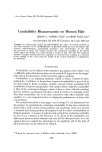
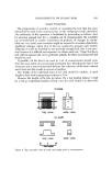
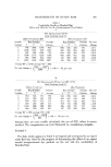
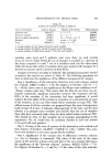
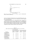
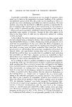
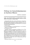
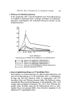
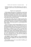
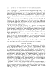
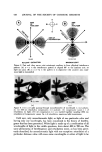
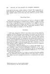
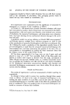
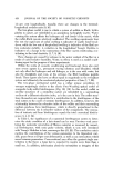
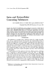
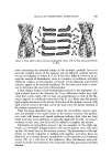
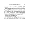
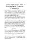
I. Soc. Cosmet. Chem., 27, 433-441 (September 1976) Intra and Extracellular Cementing Substances H. P. BADEN, M.D., L. D. LEE, Ph.D. and J. KUBILUS, Ph.D.* Presented May 1975, SCC Seminar, St. Louis, MO. Synopsis: The STRATUM CORNEUM consists of flattened compacted cornified CELLS which are filled with cross-linked FIBROUS PROTEINS. The association of the fibrous proteins with a SPECIFIC LIPID gives rise to the barrier characteristics of the epi- dermis. Stratum cornettm cells are attached to one another by desmosomes and an inter- cellular cementing substance. The latter material has been rather poorly documented and described. Recent studies concerning diseases associated with hyperkeratosis which em- ployed a keratolytic gel, have suggested that solubilization of this material can result in the loss of adherence of cells to one another. The solubilized material appears to. have unique properties, which will be characterized. INTRODUCTION' The epidermis is a complex tissue, which by means of a variety of mecha- nisms, acts as a protective barrier for the body. Our understanding of how it operates at the molecular level is gradually expanding, but some aspects have proved more difficult to investigate. This paper will deal with those factors responsible for ,maintaining the integrity of the tissue, these being the cement materials. This term is used in a very broad sense, since a number of the struc- tural components appear to play some role. We can divide these substances into materials which hold a single cell together and those which hold groups of cells together. A maior component of the epidermal cell is the a fibrous protein, which ap- pears as filaments in electronmicrographs of the skin (Fig. 1). These fila- ments are first observed in the basal layer and go through a series of changes as the cells ascend into the stratum corneum. It is thought that these 70-80 A filaments extend across the cell from one wall to another and hook on to attachment plates of desmosomes. Since it has been estimated that the basic fibrous protein has a length which is only a fraction of the width of a cell, the filaments must result from an aggregation of fibrous proteins. The fibrous pro- *Department of Dermatology, Harvard Medical School, Massachusetts General Hos- pital, Boston, Massachusetts 02114. 433

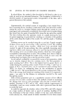
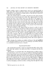
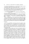
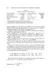
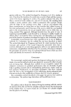
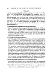
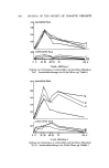
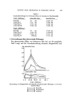
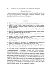
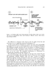
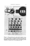
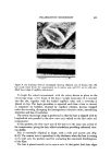
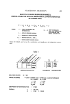
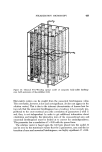
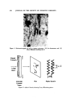
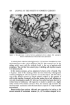
434 JOURNAL OF THE SOCIETY OF COSMETIC CHEMISTS Figure 1. Electromicrogral•h of cell in stratum s10inosum: (D) its desmosome and (F) shows filaments inserting into it Aligned Filaments X-Ray Beam ß 9.8[ ¾. Stretched Film Alpha Keratin Oriented Tissue Figure 2. ,, helix of keratin showing X-ray diffraction pattern
Purchased for the exclusive use of nofirst nolast (unknown)
From: SCC Media Library & Resource Center (library.scconline.org)