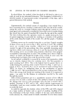
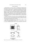
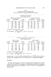
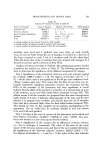
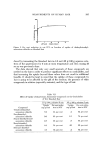
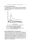
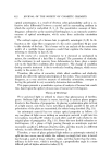
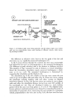
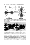
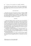
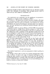
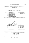
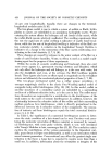
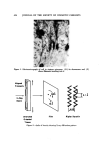
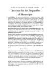
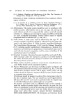
CELLULAR CEMENTING SUBSTANCES 437 ,' Figure 4. Polar lipicl is shown between polypepticle chains with its long axis perpenclicular to fibre exists concerning the chemical nature of this material, probably has as its basis the complex nature of the material and the different methods used by several investigators to isolate it (7, 8). It has been difficult, however, to ac- cept this concept of keratohyalin, since in a number of conditions, including ichthyosis vulgaris, no keratohyalin is formed, yet the filaments and stratum corneum appear to be normally stabilized. We feel that more work is neces- sary to determine the exact role of keratohyalin. A final unique feature of the keratinization process is the deposition of a lipid material between the filaments (9). X-ray diffraction studies have indi- cated that a polar lipid, with its long axis perpendicular to that of the fila- ments, appears as cornification procedes (Fig. 4). It is likely that this protein lipid complex functions as the barrier. Extraction of the stratum corneum with lipid solvents removes the lipid, and at the same time, the barrier function of the stratum corneum is lost. What has been described may be called the intracellular cement mate- rials and probably is the major barrier of the stratum corneum. Our prelimi- nary work with human and animal epidermis indicates that, what has beer• found in bovine snout epidermis, is generally applicable to both. As research continues in this area, new facts will be added to complete the picture. Information on intercellular cement is far less complete. In the viable epi- dermis, no irreversible linkage between cells can be present, since cells move up from the basal layer to the stratum corneum. The desmosomes of the epi- dermis are clearly important in holding cells together, and when these are disturbed, as in certain diseases, acantholysis or cell separation and blistering occurs (10). As a cell rises in the epidermis, these attachments must con- stantly be broken and reformed. The mechanism for this process has not been clarified.

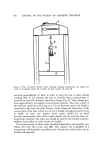
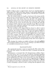
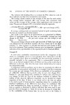
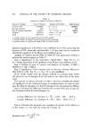
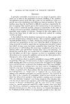
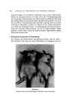
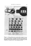
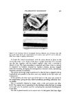
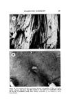
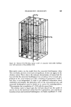
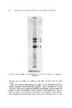
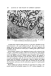
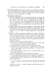
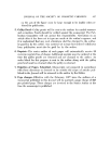
438 JOURNAL OF THE SOCIETY OF COSMETIC CHEMISTS Figure 5. The glycocalyx is shown between epidermal cells in culture. The material is stained with Ruthensum red and has fuzzy appearance A carbohydrate material called glycocalyx (11) has been described as coat- ing keratinocytes in the viable epidermis (Fig. 5). This material may be the antigen which reacts with the antibody found in the sera of patients with pemphigus. The role that this materia'l plays in holding cells together remains to be demonstrated. In the stratum corneum, a firm attachment between cells is formed. This is in part, a result of the stacking (which has been observed), which permits careful overlapping of cell and maximum use of cell surfaces (12). The thick- ness of the stratum corneum is almost certainly related to the capacity of cells to stick together. Eventually, at the skin surface, loss of cell adhesion oc- curs and desquamation results. By inference from what has been observed in certain forms of ichthyosis, cell separation is easier to achieve at a higher water content of the stratum corneum. Thus, the common type of winter dry skin frequently ameliorates when the individual is exposed to a high humidity environment. Recent studies have perhaps indicated new approaches for looking at the cement material of the stratum corneum (13, 14). In studies designed to im-
Purchased for the exclusive use of nofirst nolast (unknown)
From: SCC Media Library & Resource Center (library.scconline.org)