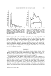
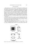
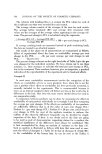
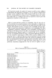
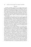
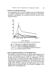
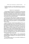
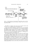
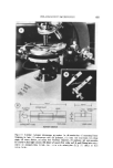
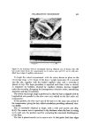
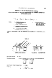
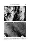
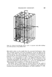
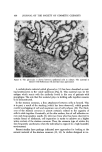
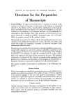
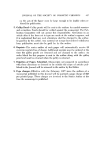
CELLULAR CEMENTING SUBSTANCES 439 prove the therapy of ichthyosis, it was discovered that mixtures of propylene glycol in the 40 to 80 per cent range in water, under plastic occlusive dress- ings, resulted in rapid shedding of the stratum corneum. This appeared to be true for ichthyosis vulgaris and sex-linked ichthyosis. A marked increase in the effectiveness of the treatment resu]te(! from the addition of salicylic acid. A gel containing salicylic acid and propylene glycol, which worked quite effec- tively has been finally developed. Use of this preparation under occlusive plastic dressings overnight resulted in rapid and dralnatic loss of the thick- ened stratum corneum. Not only could this effect be observed in ichthyosis vulgaris and sex-linked ichthyosis, but in lameIlar ichthyosis as well (Fig. 6). One could observe true keratolysis that is separation of the stratum corneuln in sheets, which could be removed by rubbing the skin when the dressings were removed. This action of propylene glycol and salicylic acid is not peculiar to the stratum corneum of ichthyosis, but can also be observed in hyperkeratosis as- Figure 6. Appearance of skin of individual with lamellar ichthyosis after treatment with salicylic acid propylene glycol gel: untreated on right and treated on ,left

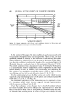
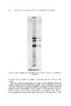
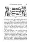
440 JOURNAL OF THE SOCIETY OF COSMETIC CHEMISTS sociated with psoriasis and eczema (15). It effects the stratum corneum of all body surfaces including the palms and soles. This can also be observed with normal skin, indicating that factors involved in thickening of the stratum cor- neum may be an exaggeration of normal mechanisms for holding the stratum corneum together. The nature of the cementing substances in the stratum corneum remains to be demonstrated. Although they are difiqcult to visualize, still the desmosomes are present and may have been modified to become very resistant cross-links by dessication. In addition, intercellular material has been described as ap- pearing above the granular layer. This material has been poorly defined, but has been proposed by some authors as a cementing substance. It is not known how this material relates to the glycocalyx in the viable layers of the epider- mis, and some have suggested that it comes from the membrane coating gran- ules. Finally, as yet unrecognized materials may play a major role. A firm fact of some significance is that hair and nail must have quite dif- ferent mechanisms for holding cells together. The propylene glycol solutions and propylene glycol and salicylic acid gel do not cause keratolysis of nail and hair even after prolonged use. If mechanisms similar to stratum corncure were involved in holding cells together, these tissues would not be as resistant. A reasonable approach to this problem is to treat stratutn corneum with agents known to produce keratolysis and to determine the nature of the solu- bilized products. Recent work has begun in our laboratory, which uses solu- tions of propylene glycol and salicylic acid. For technical reasons, materials with molecular weights below several thousand are not amenable to investi- gation in our preliminary study. However, we have identified solubilized polypeptides in the molecular weight range 5,000 to 15,000 using electro- phoretic techniques. Identification of their chemical composition is in pro- gress, and these may give clues to importance in cell cement. A½•ZNOWL•.r•CM• NTS We would like to thank S. I. Roth and W. H. Clark, Jr., for Fig. 1, which appeared as Fig. 4 in The Epidermis, W. Montagna and W. Lobitz, Eds., 1964, p. 311 and P. Fritsch, K. Wolff, and H. H/•ningsman, for Fig. 5, which appeared as Fig. i in I. Invest. Dermatol., 64, 31 (1975). REFERENCES (1) A. G. Matoltsy, Biolog!I o[ the Skin and Hair Growth, A. G. Lyne and B. F. Short, Eds., Elsevier, Amsterdam, 1965, P. 291. (2) K. M. Rude]i, The proteins of the mammalian epidermis, in Advances in Protein Chemistry, M. L. Anson, K. Barley, and J. T. Edsall, Eds., Academic Press, Inc., New York, Pp. 253-90. (3) H. P. Baden, L. A. Goldsmith, and B. Fleming, Polypeptide composition of epider- mal prekeratin, Biochim. Biophys. Acta, 317, 303-11 (1973). (4) H. P. Baden and L. Bonar, The a fibrous proteins of epidermis, I. Invest. Dermatol., 51, 478-83 (1968). (5) H. P. Baden and L. A. Goldsmith, Changes in the a fivrous protein during epider- mal keratinization, Acta Dermatovener, 51, 321-26 (1971).
Purchased for the exclusive use of nofirst nolast (unknown)
From: SCC Media Library & Resource Center (library.scconline.org)