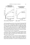
SCANNING ELECTRON MICROSCOPY 511 the relatively subtle changes brought about by the application of a cosmetic product. In fact, even an oily material applied to the surface of a biopsy sam- ple will tend to diffuse away into the embedding material and not remain on the sample. Hence, for all of these reasons, SEM tends to be the tool of choice for most skin studies. Our first SEM studies on human skin did involve biopsy samples, both ani- mal as well as human. We learned quite quickly that the severe dehydration effects of the ultra high vacuum within the SEM left the sample so severely shrunken, distorted, and changed that any relationship between reality and what we were seeing would have certainly been fortuitous. Even some of the newer and more sophisticated methods of sample preparation, such as critical point drying or freeze drying, were not particularly better. Consequently, we have concentrated virtually all of our efforts toward procedures employing skin replicas, which have the advantage that we cannot only characterize by SEM the in vivo state of an area of stratum corncure, but we can also follow the same identical area in the micrographs as a function of time, in a sense, like a high magnification form of time lapse photography. In this way, 'we can follow the effects of a product as a function of time, and evaluate the ef- ficacy of this product relative to a control. Surely, we •vere not the first to make replicas of human skin. Facq (9, 10) and Berstein (11, 12) learned back in the late 1960s that normal silicone resin, either of the RTV-11 type* or even the impression materials used in dentistry, would make "acceptable" (relatively free of artifacts) replicas of the top surface of the skin (and, hence, also the top surface of the stratum comeurn). Once having obtained the "impression" or "negative" as we prefer to call it, the negative is itself replicated, yielding a "positive" replica of the skin surface. There was also a problem with artifacts in the replicas, such as air bubbles, which tended to limit, in certain respects, the utility of these original efforts. Unfortunately, artifacts were not the only drawbacks t ø the methods origi- nally used. First, was the criticism that too small an area was being studied how was it known that this small area was indeed representative of that area of stratum corncure as a whole? In addition, the enormous heterogeneities present from point to point on the stratum corncure were such that one could almost prove or disprove anything by a careful selection of the area. Our approach was to develop a procedure that would enable us to do the following. 1. produce high resolution (virtually) artifact-free negative replicas 2. convert the negative replicas to high resolution positive replicas *General Electric Company, Wateroeord, N.Y. 12188.

512 JOURNAL OF THE SOCIETY OF COSMETIC CHEMISTS 3. photograph the same identical areas, before and after treatment, as a function of time, and by following the same identical area, avoid some of the problems of hetcrogeneity from point to point 4. make large montages, rather than randomly selected pictures so it would be obvious a large enough area was being photographed 5. quantify the resulting images either manually or through the use of an image analyzing computer (such as a Quantimet) 6. correlate the results, quantitatively as well as qualitatively, with known clinical conditions. The purpose of this paper, therefore, is to describe some of the methods we have developed for this purpose, to present the clinical basis for justifying this approach, and to show how the resulting data is presently being used to dem- onstrate efficacy and document advertising claims. EXPERI1VIENTAL A. Preparation o• Negative Replicas RTV-11 sfiicon resin is acceptable for making the negative replicas, al- though we find that better results are obtained with the high molecular weight fraction removed and when an absolute minimum of catalyst is used to setup the resin. Certain silicone-compatible surface active agents (such as "Aero- sol OT,"*) seem to impart better release characteristics and may have their maximum benefit when replicating particularly rough skin (such as psoriatie subiects). B. Conversion o[ Negative Replicas to Positive Replicas Almost any thermoplastic resin can be used for this purpose, but we prefer fine spray-dried or asopolymerized polyolefin "bead" polymer. After the posi- tive replica is completed, it is metallized to make the replica surface conduc- tive to prevent sample charging in the electron beam. C. Preparation o[ Replica Cross-Sections For replica cross-section work, the positive replicas are diamond knife- trimmed, using a duPont/Sorvall MT2-B Diamond Knife Ultramicrotome.t The knife speed and angle must be adjusted for minimum chatter and thin- nest sections (even though the sections are not important) as these are the conditions for obtaining the best undistorted and unsmeared cross-sections. Unfortunately, the sorer resins yield the best detail in the positives hence, we sometimes must do the sectioning in a cryostage at liquid nitrogen tem- *American Cyanamid Co., Stamford, CT. rE. I. dupont de Nemours and Co., Inc., Wilmington, DE 19898.
Purchased for the exclusive use of nofirst nolast (unknown)
From: SCC Media Library & Resource Center (library.scconline.org)