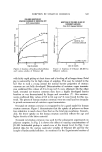
SCANNING ELECTRON MICROSCOPY 513 peratures as a means of minimizing smearing and preserving the cross-section- al edge. We have not been successful using glass knives for this purpose. D.SEM The work described in this paper was done on a JEOL Model #JSM-U3 SEM.* We have found this equipment to be particularly well-suited for this type of work for several reasons: (1) it can be modified without too much dif- ficulty to provide a virtually distortion-free image in the 15 to 20 X range, an absolute necessity for putting together distortion free montages (2) it has an extremely flexible goniometer stage, with all of the degrees of freedom neces- sary to relocate the same identical areas of a replica without exceptional dif- ficulty and (3) the electronic stability of the instrument is such that micro- graphs can be taken without discernable drift in brightness or contrast, an ex- tremely important feature xvhen montages are being made. The most important aspect of our procedure is that every successive replica in a series is done on the very same identical area, same tilt, same magnifi- cation, in short, every aspect of the images are identical. We have eliminated any chance that a particular feature of the results might be due to the way the microscopy was done rather than due to real changes on the skin sur- face. This also means, therefore, that all microscopy, which is done generally in the form of large montages, is clearly from the same identical areas and this fact is so obvious, that even a nonmicroscopy-oriented observer would realize it. E. Quantitative Image Analysis All image analysis was done on a Quantimet 720B Image Analysing Com- puter equipped with the automatic detector, optical and epidiascope input modes, and classifier-collector modules with direct teletype output. RESULTS An example of what one can typically obtain is shown on Fig. 1. This is an area from the back of the hand of a young female subject in her mid-twenties, and the microscopy was originally done as a montage at 100 X from the scanning electron microscope (but which was reduced for publication). The original individual fields that make up the montage can still be seen. The "hill and valley" type of topography is "normal," particularly for young- er subjects. The one major exception is what is sometimes called the "cobble- stone" texture and an example of it is shown in Fig. 2. The term "cobblestone" *Jeol (USA), Inc., Medford, MA.

514 JOURNAL OF THE SOCIETY O1•"COSMETIC CHEMISTS Figure I. SEM montage taken from positive skin replica from back of hand of female subiect in mid-20s. Example of "good" skin. Scale bar represents •00 txm. texture was originally coined by D. R. Highley (13) who has four•d that this texture occurs in approximately 5 percent of the population. For studies on skin care products, we generally try to avoid using "cobblestone" subiects, be- cause the effects of cosmetic products are more difficult to discern in the final montages and the quantitative analysis is more difficult. One must be extremely careful when making both the negative as well as the positive replicas, so as not to produce artifacts. Figure 2 was actually made in mid-1972, and was one of our earliest attempts. It also contains the 3 most commonly found artifacts: (1) air bubbles in the negative replica (2) air bubbles in the positive replica and (3) a glazed area of improperly mixed replicating material (in the negative). Even our most experienced replica makers still occasionally produce these artifacts, but the goal is always to minimize their frequency of occurrence. In this way, even though some areas of a replica might be bad, we can generally find at least some areas which have replicated well in all the replicas of a sequence. Type A artifacts can be minimized by using only the freshest resin and catalyst, which are then mixed smoothly without whipping any unnecessarily large amount of air into the
Purchased for the exclusive use of nofirst nolast (unknown)
From: SCC Media Library & Resource Center (library.scconline.org)