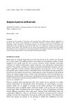
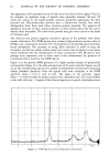
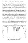
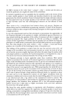
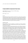
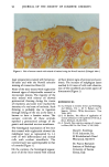
Letters to the Editor To the Editor: The disputed (1) assertion by Butcher (2) that mineral oil produces hypertrophy of the epidermis finds a measure of support in our recent study of albino white mice. Male and female Swiss albino mice were depilated on their backs (area of 2 cm 2) with a commercial depilatory cream. Small quantities of mineral oil USP (Sin- clair-Arco) were applied to depilated backs of 25 mice with sterile cotton pads while another lot of 25 mice was treated in a similar way with a triglyceride (Nesa- tol ©, Laboratori Vevy, Genoa, Italy). After 65 days of treatment (depilatory was used intermittently every 12-15 days) all the mice were beheaded and portions of the treated skin were taken aside, together with adjoining edges that were used as normal controls. The pieces of skin that were immediately fixed in 10% formalin solution were then used to prepare histo- Figure 1. Skin of mouse treated with triglyceride stained using the Novelli method. (Enlarged 100x.) 93

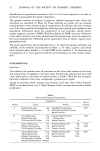
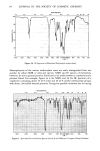
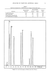
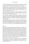
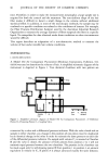
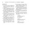
94 JOURNAL OF THE SOCIETY OF COSMETIC CHEMISTS Figure 2. Skin of mouse treated with mineral oil stained using the Novelli method. (Enlarged 100x .) logic preparations stained with hematoxy- lin-eosin and with the Novelli selective staining of connective fibers (3). None of the mice treated with triglyceride showed signs of objectivable external or necroscopic lesions. The majority of the mice treated with mineral oil showed pronounced thinning during the course of treatment, and some mice reached the intensity of a real state of cachexia. Such thinning is probably due to ingestion through licking of the oil, as the latter is known to have a laxative action. The autopic controls of these animals reported a pronounced athropy of the gastric and intestinal mucous membrane. The histological examination of the mice skin treated with triglyceride showed the malphigian layer as represented by 2-3 rows of well differentiated and normal cells with evident tonofibrils. The corneum layer was superimposable to that of controls (Figure 1). On the contrary, the histological appear- ance of all the mice treated with mineral oil have shown signs of pronounced acan- thosis. The increase of malphigian layers reached 9-10 rows of cells with dissocia- tion of the tonofibrils and some aspect of disceratosis (Figure 2). REFERENCES (1) S.J. Strianse in Cosmetic Science and Technology, 2nd Edition, Vol. 1, M. S. Balsam and E. Sagarin, Editors (Wiley-Interscience, New York, 1957). (2) E. O. Butcher, The effects of application of various substances in the epidermis of the rat,J. Invest. Dermatol., 16, 88 (1951). (3) A. Novelli, New selective staining of connect- ive fibers, Boll. Soc. Hal. Patol., 5, 145 (1958). David C. Steinberg Tri-K Industries, Inc. 99 Kinderkamack Road Westwood, NJ 07675 Dr. Franco Burlando Institute of Pathology University of Genoa Genoa, Italy
Purchased for the exclusive use of nofirst nolast (unknown)
From: SCC Media Library & Resource Center (library.scconline.org)