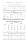
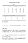
304 JOURNAL OF THE SOCIETY OF COSMETIC CHEMISTS Caprylic/capric triglyceride has been used in cosmetic products because of skin coverage properties and because it is an excellent emollient. The suppliers of this product claim that it can promote skin penetration however, no published data is available to verify these claims. Caprylic/capric triglyceride has been shown to be non-irritating (using Draize skin test and skin patch test) (29,30) and non-sensitizing (29). The purpose of this study is to compare the influence of propylene glycol and caprylic/ capric triglyceride on the in vitro percutaneous absorption and the in vivo vasoconstric- tion activity of various steroids. EXPERIMENTAL CHEMICALS Hydrocortisone 17-butyrate (Gist-Brocades, Delft, The Netherlands), desonide (S.I.R.S. Spa, Milan, Italy), hydrocortisone (Upjohn, Kalamazoo, MI), diflorasone diacetate (Upjohn, Kalamazoo, MI), propylene glycol (Dow Chemical Co., Midland, MI), caprylic/capric triglyceride (Ionelex Chemical Div., Philadelphia, PA), polysor- bate 60 (ICI Americas, Wilmington, DE), sorbitan stearate (ICI Americas, Wil- mington, DE), sodium phosphate (dibasic) 0. T. Baker Chemical Co., Phillipsburg, NJ), sodium phosphate (monobasic) and sodium chloride (Mallinckrodt, St. Louis, MO) were used as supplied. Methanol and acetonitrile were of HPLC grade 0- T. Baker Chemical Co., Phillipsburg, NJ). ANALYTICAL Apparatus. For the HPLC separation, the following instruments were used: Waters Associates Model 510 pump, Model 481 variable wavelength detector, WISP 710B autoinjector, and Data Module integrator. A reversed phase Zorbax O.D.S. (10 chromatographic column (Supelco Inc., Bellefonte, PA) was used for separation. Chromatographic conditions. Th6 composition of the mobile phase, the flow rate of the mobile phase and the wavelength used to assay the steroids are listed in Table I. The solvents were filtered and then degassed under vacuum prior to use. ANIMALS Homozygous athymic male hairless mice (Harlan Sprague Dawley, Inc., Indianapolis, IN), between the age of 42 and 56 days, were used. Table I Chromatographic Conditions Flow Rate Wavelength Steroid Mobile Phase* mL/min nm Hydrocortisone CH3OH:CH•CN:H_•O, 30:30:40 1.1 264 Hydrocortisone 17-butyrate CH3OH:CH3CN:H20, 32.5:32.5:35 1.1 264 Desonide CH3OH:CH3CN:H20, 30:30:40 1.1 264 Diflorasone Diacetate CH3OH:CH•CN:H:O, 25:25:50 1.1 264 * By volume.

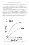
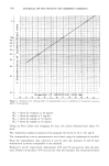
COMPARISON OF TOPICAL VEHICLES 305 FRANZ DIFFUSION CELLS (Crown Glass Co., Somervd/e, NJ) The cells are made up of the cell cap (donor) and the cell body (receiver). The cell cap was open to the air and allowed the application of a finite dose to the membrane. The volume of the cell body was 11 mL. The skin was mounted between the two ball joints, on an O-ring, using a pinch-type, ground joint clamp. The available area for diffusion was 3.14 cm 2. The epidermal side of the skin was exposed to ambient labo- ratory conditions, while the dermal side was bathed by a phosphate-buffered saline solution containing 0.02% thimerosal. Stirring of the receiver solution was accom- plished by a teflon-covered magnetic stirring bar, driven by an external magnet. The experiments were conducted at ambient temperatures (21 _+ IøC). PREPARATION OF SOLUTIONS The compositions of vehicles used in this study are given in Table II. An excessive amount of steroid was added to a 10 mL test tube containing the vehicle. The test tubes were shaken mechanically at room temperature for 24 hours. The saturated solutions were centrifuged (IEC HN-SII Centrifuge, International Equipment Co., Needham, MA) until the supernatant contained no crystals as determined by micro- scopic analysis. The crystal-free supernatant was then analyzed by HPLC for the con- centration of steroid. IN VITRO PERCUTANEOUS A•SORPTION STUDY Skin preparation and mounting. Mice were sacrificed by severing the spinal cord. A rectangular section of the abdominal skin was excised from the animal with surgical scissors. Adhering fat and other visceral debris were removed carefully from the under surface with tweezers. The skin was then mounted between the two cells and the receiver side was filled with the phosphate-buffered saline solution. Permeation. Following mounting, at time zero, 100 }xL of test solution was placed on the epidermal surface. One hundred [xL was sufficient to spread across the entire epi- dermal surface. Samples were withdrawn from the receiver compartments at 6, 24, 30, 48, and 72 hours and were analyzed by HPLC. Area under the curve was used to quantirate drug concentration in the receiver solution. Each experiment was performed at least in triplicate. The permeability data was plotted using the total amount of drug penetrated as a Table II Compositions of Vehicles Used for In Vitro and In Vivo Testing Parts by Volume Ingredient Vehicle No. 1 Vehicle No. 2 Sorbitan Stearate 1 1 Polysorbate 60 3 3 Propylene Glycol 10 -- Caprylic/Capric Triglyceride -- 10 Water 70 70
Purchased for the exclusive use of nofirst nolast (unknown)
From: SCC Media Library & Resource Center (library.scconline.org)