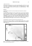
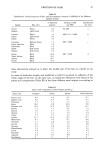
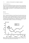
j. Soc. Cosmet. Chem., 40, 101-107 (March/April 1989) The role of antioxidants in skin immune reactions: The use of flow cytometry to determine alterations in la-positive epidermal cells in allergic contact dermatitis LAWRENCE A. RHEINS,* RICHARD A. MORAVEC, MICHAEL L. NORDLUND, LINDA S. TRINKLE, and JAMES J. NORDLUND, Department of Dermatology, University of Cincinnati College of Medicine, Cincinnati, OH 45267. Synopsis Earlier experiments from our laboratory revealed that common parasubstituted phenolic compounds such as monobenzyl ether of hydroquinone (MBEH), butylated hydroxytoluene (BHT), and butylated hydroxyani- sole (BHA), ubiquitous compounds found in medications, cosmetics, foods, and various industrial products, can alter Ia+ Langerhans cells (epidermal macrophages) and epidermal immune responsiveness (i.e., exacerbate allergic contact dermatitis) when topically applied to the epidermis of various strains of mice. The current experiments demonstrated that a precise and sensitive instrumental approach, fluorescent activated cell sorting (FACS), could be used to quantify changes more precisely in the expression of the membrane-bound Ia antigen found on epidermal antigen-presenting-Langerhans cells during contact hyper- sensitivity reactions. Further, this technique can be used to screen potential vehicle and/or medicament allergens as likely sources for allergic contact dermatitis at home and in the work place. INTRODUCTION Earlier studies from our laboratory have documented that topical applications of common antioxidants such as monobenzyl ether of hydroquinone (MBEH), butylated hydroxyanisole (BHA), and butylated hydroxytoluene (BHT), ubiquitous compounds found in medications, cosmetics, food, and industrial rubber products, can alter the contact hypersensitivity responsiveness (CHS) (e.g., allergic contact dermatitis, ACD) in various mice strains when epicutaneously applied (1). These compounds, when topi- cally applied for a short period of time (i.e., daily for five days) to various epidermal sites, can alter the density (cells/ram 2) of Ia + (immune associated antigen)/ATPase + Langerhans cells (LC) (epidermal immune macrophages). Further, this increase or de- crease in identifiable Langerhans cells could be correlated with alterations in CHS re- sponsiveness following sensitization and challenge with common allergens/haptens (i.e., dinitrofiuorobenzene, oxazalone) (1). Other studies suggest that the mechanism responsible for the alteration of Ia + Langerhans cell density and functional CHS reactivity following topical antioxidant treatment may be mediated via arachidonic acid (AA) and/or its metabo- lites (i.e., prostaglandins, thromboxanes, leukotrienes)(1,2). * Reprint requests should be sent to: Lawrence A. Rheins Ph.D., Procter & Gamble Co., Ivorydale Tech- nical Center, Cincinnati, Ohio 45217 101

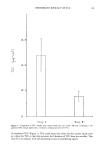
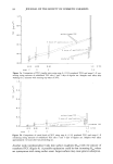
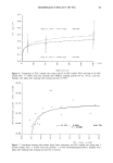
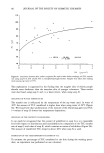
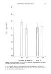
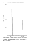
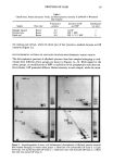
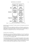
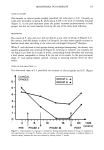
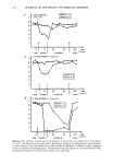
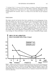
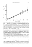
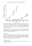
102 JOURNAL OF THE SOCIETY OF COSMETIC CHEMISTS The current study was designed to examine, using more precise methodology (flow cytometry), the density of the Class II major histocompatability (MHC) Ia antigen on epidermal immune cells that had been previously treated in intact epidermis with various antioxidants or a biologically relevant molecule, arachidonic acid. It has been previously documented that the density of the Ia antigen membrane marker correlates with the intensity of epidermal immune reactivity (e.g., allergic contact dermatitis (1,3,5). We suggest that the described methodology may be useful as a sensitive screening technique for ascertaining whether common and/or new antioxidants used in medications and/or cosmetics may exacerbate common immune-mediated dermatoses such as allergic contact dermatitis. MATERIALS AND METHODS Four-to-six-week-old male DBA/2 d and C57BL/6 b mice were obtained from Jackson Laboratories (Bar Harbor, Maine) and were housed in University facilities, four to six per cage with free access to water and Purina Mouse Chow © . The animals were exposed to an ambient 12-hour light:dark photoperiod. The experiments were performed at least twice and produced similar results. IN VIVO EXPOSURE TO MBEH AND AA DBA/2 mice were treated topically with either 50 }zl of 20% MBEH in 95% ETOH or 0.05% and 1.0% AA in DMSO:H20 (1:1) applied to the dorsal pinnal epidermis daily for five days. Control animals were topically treated with 95% ETOH diluent or the DMSO:H20 (1:1) diluent. On day 6, treated and diluent control ears were surgically excised and epidermal suspensions were prepared and subjected to fluorescent-activated cell sorting analysis as described by others (6,8,9) with modifications. IN VITRO EXPOSURE TO AA, BHT, BHA 4 x 10⁵ viable unenriched epidermal cell suspensions were prepared and plated in 24-well Linbro polystyrene tissue culture plates. To the unenriched epidermal cells, various concentrations of AA, BHT, and BHA were added in complete RPMI 1640 cell culture media supplemented with 1.5 }zg/ml of indomethacin. A series of pilot experi- ments was performed at various concentrations to determine the in vitro effects of the specific phenols BHT, BHA, and AA. The maximum biological effects were produced by the following solutions: BHT, 2.5 !zg/ml BHA, 5 !zg/ml and AA, 66 ng/ml and 666 ng/ml therefore, these concentrations were used throughout the studies. Following 48 hours of incubation at 37øC in 5% CO2, the cells were harvested, washed in com- plete 1640 RPMI medium, and incubated with appropriate primary and secondary fluorochrome-conjugated antibodies at 4øC (8,9). Treated and control epidermal cell preparations were then subjected to FACS analysis and the density of Ia expression quantified. To ensure that the cells were Langerhans cells and not keratinocytes which can also express the Ia membrane antigen in allergic contact dermatitis, unenriched epidermal cell suspensions were double stained with the Ia and Fc receptor antigens. The antigens were then conjugated with appropriate secondary antibodies, specifically fluorescein isothiocyanate and streptavidin phycoerythrin red.
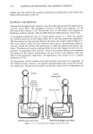
Purchased for the exclusive use of nofirst nolast (unknown)
From: SCC Media Library & Resource Center (library.scconline.org)