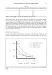
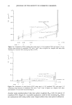
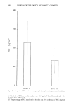
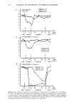
J. Soc. Cosmet. Chem., 40, 119-125 (March/April 1989) Skin penetration of vitamins C and E K. TOJO and A. C. LEE, Department of Pharmaceutics, College of Pharmacy, Rutgers-The State University of New Jersey, Busch Campus, Piscataway, NJ 08855-0789. Received February 23, 1988. Synopsis The skin penetration profile of vitamin C determined by a H'PLC assay procedure agreed approximately with that determined by liquid scintillation counting using the radiolabeled vitamin. This finding indi- cates that only a small amount of vitamin C is bioconverted to its metabolites in the hairless mouse skin. However, the lag time in the penetration profile assayed by HPLC was appreciably shorter than that determined by radioactivity counting. This finding suggests that the tissue vitamin C distributed initially either endogenously or exogenously might diffuse into the receptor solution during the transient period of penetration. The penetration profile of vitamin E determined by HPLC differed markedly from that determined by radioactivity counting. During about 48 hours after the onset of the penetration experiment, the skin penetration of vitamin E remained almost negligible. Beyond that time, however, vitamin E appeared gradually in the receptor solution. By assuming first order kinetics of vitamin E bioconversion in the viable skin and the exponential decay law with respect to enzyme deactivation under in vitro conditions, the time course of the cumulative appearance of the vitamins after skin penetration was described on the basis of the bilayer diffusion/bioconversion model. INTRODUCTION During the last two decades, many researchers have revealed various biochemical func- tions of vitamins C (ascorbic acid) and E (ot-tocopherol) (1,2,3). The use of these vi- tamins is unquestionably important not only for maintaining normal body metabolism but for preserving healthy skin. It was reported that vitamins C and E protect synergis- tically against the peroxidation of membrane lipid (4). Various cosmetic formulations for skin care contain vitamins C or E as an active ingre- dient. However, the skin permeation of these vitamins has not been elucidated yet. We still lack much of the information we need to understand the percutaneous absorption of these vitamins. In this communication, we have investigated the skin penetration of vitamins C and E using hairless mouse skin in vitro. The penetration profiles (time course of the cumula- tive amount of vitamin penetrated) for both radiolabeled and nonradiolabeled vitamins were determined using either HPLC or a liquid scintillation counter. The penetration 119

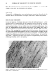
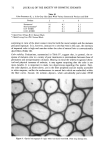
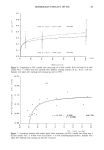
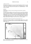
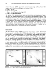
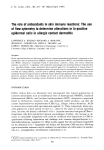
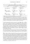
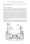
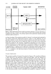
120 JOURNAL OF THE SOCIETY OF COSMETIC CHEMISTS profiles were then analyzed by a dynamic mathematical model based on the bilayer skin diffusion/bioconversion model (5). MATERIALS AND METHODS Vitamin E (o•-tocopherol) and vitamin C (ascorbic acid) were provided by Sigma Chem- icals (St. Louis, MO). The radiolabeled vitamin E ((3H)2-3,4o•-tocopherol, 10-40 mCi/mmol) and vitamin C L-[1-•4C]-ascorbic acid, 10 mCi/mmol) were supplied by Hoffmann-La Roche (Nutley, N J) and NEN Research (Massachusetts), respectively. A full-thickness abdominal skin of a female hairless mouse (5-7 weeks old, Jackson Lab. HRS/J strain) was excised freshly before the in vitro skin penetration experiment. The skin sample was then mounted between the donor and receptor halfcells (Figure 1). The in vitro system, which has been calibrated with respect to hydrodynamic character- istics (6), assures the intrinsic skin penetration (7) under the present experimental con- dition. The donor and receptor solutions (Table I) were then charged in each cell com- partment. At appropriate time intervals, 30 }xl of the receptor solution was withdrawn and assayed for the vitamin concentration. Six sets of the in vitro diffusion cells were used in each penetration experiment. All experiments were carried out at a constant temperature (37øC). The degradation of both vitamins in the donor solution was found to be negligible. In the receptor solution, however, the vitamins degraded appreciably during the penetra- tion experiment. The penetration profile was therefore corrected for the degradation by ( ,••Stopper • St•rnng [• j (10 mm dia.,
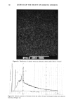
Platform 4 mm hgt ) .., '•"-• / Slar-hne•d 35ml .... capacity Filling & Sampling Port Skin lIGHT SEAL DONOR (Left) HALF-CELL RECEPTOR (Righi) HALF-CELL Figure 1. In vitro skin penetration apparatus used in this study.
Purchased for the exclusive use of nofirst nolast (unknown)
From: SCC Media Library & Resource Center (library.scconline.org)