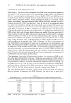
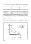
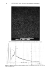
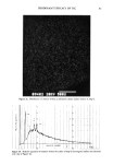
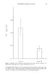
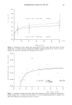
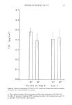
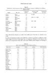
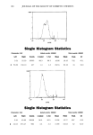
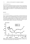
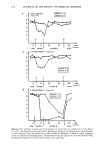
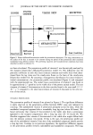
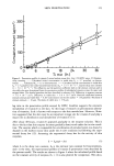
SKIN PENETRATION 121 Table I The Donor and Receptor Solutions Used in the In Vitro Skin Penetration Study Drug in donor Donor solution Receptor solution Vitamin C Non-radiolabeled vitamin C 50% glycerin aq. solution 12.11 -+ 0.86 mg/ml and Radiolabeled •4C-vitamin C 5.93 --- 0.25 X 10⁵ DPM/ml in 50% glycerin aq. solution. Non-radiolabeled vitamin E 13.81 -+ 0.51mg/ml and Radiolabeled 3H-vitamin E 1.23 + 0.09 X 10⁵ DPM/ml in silicone fluid (DC360, 20 cp) Vitamin E 5 mM Tween-80 aq. solution the procedure described previously (8). The details of HPLC assay procedure for each vitamin were also reported previously (8). The radioactivity of radiolabeled vitamins was measured by a liquid scintillation counter. The degradation of vitamins during skin penetration is not taken into account in this assay procedure. The radioactivity in the donor solution was 1.2 X 10 5 DPM/ml and 5.9 X 10 5 DPM/ml for vitamins E and C, respectively. During the entire period of penetration experiment, the receptor solution was maintained under a sink condition for both radiolabeled and nonlabeled vitamins. The penetration profiles (time course of the cumulative amount of vitamin penetrated) were described by the bilayer diffusion/bioconversion model for percutaneous absorption (5) (Figure 2). RESULTS AND DISCUSSION VITAMIN C PENETRATION The penetration profile (cumulative amount) of vitamin C determined by either HPLC or radioactivity counting is shown in Figure 3. After about nine hours, the penetration profile for both radiolabeled and nonlabeled vitamins reached a steady state. The rate of steady-state penetration under the HPLC assay condition evaluated from the slope of the linear portion of the profile was close to, but slightly lower than, that measured by radioactivity counting. This finding indicates that vitamin C was not metabolized in the skin to a significant degree. However, the time-lag, which is defined as the time intercept of the linear portion of the penetration profile, was appreciably shorter for HPLC assay (1.3 h) than that determined by radioactivity counting (3.9 h). We re- ported previously that vitamin C quickly appeared in the receptor solution after a pro- vitamin bioconversion in the hairless mouse skin (8). The present finding, as well as our previous one, suggests that the initial tissue concentration of either endogenous or exogenous vitamin C may cause a bursting effect immediately after the onset of the penetration experiment. The tissue vitamin C in the hairless mouse skin (HRS/J strain) was recently demonstrated by Buettner et al. (9), although the concentration level has

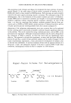
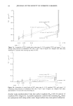
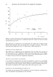
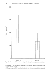
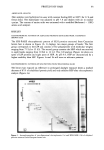
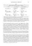
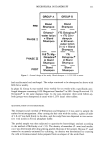
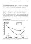
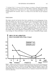
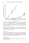
122 JOURNAL OF THE SOCIETY OF COSMETIC CHEMISTS STRATUM DONOR CORNEUM VIABLE SKIN RECEPTOR I I I I I I I DRUG DRUG Reservoir Diffusion Barrier METABOLITES Sink Condition Figure 2. Bilayer diffusion/bioconversion model for percutaneous absorption. The drug concentration on the surface of the skin is assumed to be constant during the period of skin penetration (skin-controlled transdermal drug delivery system). The governing equations and boundary/initial conditions have been described in detail in reference (5). not been elucidated. The penetration profile of vitamin C was theoretically analyzed by the dynamic bilayer-skin diffusion/bioconversion model (5). The diffusivity and the partition coefficient in each skin layer (stratum corneum and viable skin) were deter- mined from the lag times and the steady-state fluxes on the basis of the steady-state bilayer skin model (10). By assuming a homogeneous distribution for tissue vitamin C (initial concentration), the penetration profile was simulated (Figure 3) and compared with the experimental one. The initial tissue concentration of vitamin C was found to be approximately 2.7 }xmol/ml in the viable skin of the present animal model. The rate constant of vitamin C bioconversion in the skin was also found to be very small (1.5 ( 10 -5 s-1) compared to the skin bioconversion of vitamin E discussed in the next sec- tion of this article. VITAMIN E PENETRATION The penetration profiles of vitamin E are plotted in Figure 4. The significant difference is easily observed in the penetration profiles between HPLC assay and radioactivity counting. The radiolabeled vitamin E penetrated promptly across the skin, while the nonlabeled compound appeared after a remarkably long lag time (about 48 hours). We found previously that vitamin E bioconverted in the viable skin from a provitamin appeared in the receptor compartment after a long lag time (24-36 hours) (8). We therefore suggested that vitamin E bioconverted in the viable skin might diffuse back into the stratum corneum very slowly. If this is the case, the penetration profile not only of the radiolabeled vitamin E but also of the nonlabeled compound should provide a long lag time. Contrary to our previous hypothesis, the significant difference appears in the penetration profiles between radiolabeled and nonlabeled compounds. The long
Purchased for the exclusive use of nofirst nolast (unknown)
From: SCC Media Library & Resource Center (library.scconline.org)