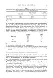
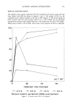
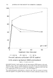
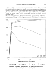
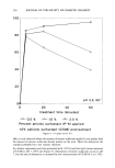
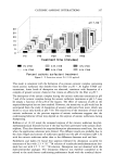
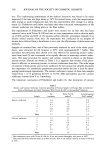
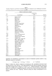
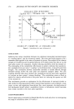
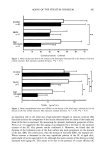
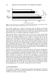
IN VITRO EFFECT OF WHITENING COSMETICS 367 on mouse hair follicle melanocytes. Topically applied 2% linoleic acid markedly sup- pressed hair growth, but the depigmenting effect was less significant (data not shown). These findings suggest that linoleic acid inhibits melanin production by its cytotoxic action. Arbutin dose-dependently reduced tyrosinase activity per well at concentrations below 1.0 mM. The amount of melanin was reduced to 75% by arbutin. The effect of arbutin was about 1/100 that of hydroquinone. Arbutin can also act as a good substrate for tyrosinase, similar to hydroquinone the effectiveness of these drugs as depigmenting agents may be related to their ability to act as substrates for tyrosinase. The depig- menting effect of a milky lotion containing 3% arbutin was tested on some forty individuals suffering UV irradiation on the inner side of the upper arm. The lotion was applied three times daily. After seven days, skin pigmentation was significantly inhib- ited compared with placebo lotion (20). Kojic acid markedly inactivated isolated tyrosinase by chelation (1). In cultured human melanocytes, tyrosinase activity per well was slightly reduced at the concentration range between 0.1 mM and 0.5 mM but was rapidly dose-dependently reduced at higher concentrations. At lower concentrations, a dose-dependent decrease was not observed. These findings suggest that the inhibitory effect of kojic acid on tyrosinase activity in the cell culture system is smaller than that of arbutin at concentrations that do not affect cell viability, even though marked inactivation was observed in isolated tyrosinase. Nair et al., using Yucatan minipigs as an assay in vivo (21), reported that kojic acid topically applied for 12 weeks resulted in no activity. On the contrary, Mishima et al. reported that topically applied kojic acid prevented artificial pigmentation in humans by irra- diation with UV (22). The discrepancy between these results may be ascribed to the difference of species or the method of in vivo testing. Further investigations are needed to clarify this. In ascorbic acid-treated cells, tyrosinase activity per well was slightly reduced at final concentrations between 0.05 mM and 0.50 mM but rapidly dose-dependently reduced at higher concentrations. Ascorbic acid was oxidized rapidly in the aqueous phase, with loss of activity in time and very limited transcellular potency owing to its being hydrophilic. Some ascorbic acid derivatives were considered stable and transcutaneous with regard to antipigmenting function (23,24). Magnesium ascorbic acid phosphate, which is a stable ascorbic acid derivative, prevented erythema and postinflammatory hyperpigmentation following UV irradiation in humans (23). Lipophilic ascorbic acid derivatives also prevent freckles and melanin spots on the skin (24). Ascorbic acid is a potent antioxidant in addition to its anti-enzymatic properties (23,26), it may prevent melanin synthesis by suppressing inflammation (25) and by inhibiting the auto- oxidation of dopa and dopaquinone. These results suggest that, to clarify those effects not ascribed to cytotoxicity, assays for both tyrosinase activity and cell viability in human melanocyte cultures are necessary to evaluate the depigmenting action. As whitening cosmetics are usually used daily, if the depigmenting effect is caused by cytotoxicity, irreversible hypopigmentation will occur somewhat in the skin or hair. In this assay system, arbutin inhibits melanin production by reducing tyrosinase activity, not by non-specific cytotoxicity, and the depigmenting action of arbutin is stronger than that of kojic acid or ascorbic acid. However, the end exposure site for whitening cosmetics will usually be intact skin. We need to consider both in vitro and in vivo effects.

368 JOURNAL OF THE SOCIETY OF COSMETIC CHEMISTS REFERENCES (1) Y. Mishima, S. Hatta, Y. Ohyama, and M. Inazu, Induction of melanogenesis suppression: Cellular pharmacology and mode of differential action, Pigment Cell Research, 1, 367-374 (1988). (2) S. Shono and K. Toda, "The Effect of Fatty Acids on Tyrosinase Activity," in Phenotypic Expression in Pigment Cell, M. Seiji, Eds. (The University of Tokyo Press, Tokyo, 1981), pp. 263-268. (3) Y. Tomira, K. Maeda, and H. Tagami. Mechanisms for hyperpigmentation in postinflammatory pigmentation, urticaria pigmentation and sunburn, Dermatologica, 179 (suppl. 1), 49-53 (1989). (4) S.S. Bleehen, Skin bleaching preparations, J. Soc. Cosmet. Chem., 28, 407-412 (1977). (5) K. A. Arndt and T. B. Fitzpatrick, Topical use of hydroquinone as a depigmenting agent, .lAMA, 194, 117-119 (1965). (6) C. B. Denton, A. B. Lerner, and T. B. Fitzpatrick, Inhibition of melanin formation by chemical agents,J. Invest. Dermatol., 18, 119-135 (1952). (7) S.S. Bleehen, M. A. Pathak, Y. Hori, and T. B. Fitzpatrick, Depigmentation of skin with 4-iso- propylcatechole, mercaptoamines, and other compounds, J. Invest. Dermatol., 50, 103-117 (1968). (8) Y. M. Chem and W. Chavin, "Hydroquinone Activation and Inhibition of Tyrosinase," in Pigment Cell, V. Riley, Ed. (Karger, Basel, 1976), Vol. 3, pp. 105-112. (9) S. Passi and M. Nazzaro-Porro, Molecular basis of substrate and inhibitory specificity of tyrosinase, Br. J. Dermatol., 104, 659-665 (1981). (10) K. Jimbow, H. Obata, M. A. Pathal, and T. B. Fitzpatrick, Mechanism of depigmentation by hydroquinone,J. Invest. Dermatol., 62, 436-439 (1974). (11) K. B. Penny, C.J. Smith, and J. C. Allen, Depigmenting action of hydroquinone depends on disruption of fundamental cell processes, J. Invest. Dermatol., 82, 308-310 (1984). (12) C. J. Smith, K. B. O'Hare, and J. C. Allen, Selective cytotoxicity of hydroquinone for melanocyte- derived cells is mediated by tyrosinase activity but independent of melanin content, Pigment Cell Research, 1, 386-389 (1988). (13) T. Mosmann, Rapid colorimetric assay for cellular growth and survival: Application to proliferation and cytotoxicity assays, J. Immunol. Methods, 65, 55-63 (1983). (14) A. Oikawa and M. Nakayasu, Quantitative measurement of melanin as tyrosinase equivalents and as weight of purified melanin, Yale. J. Bid. Med., 46, 500-507 (1973) (15) M. Eisinger and O. Marko, Selective proliferation of normal human melanocytes in vitro in the presence of phorbol ester and cholera toxin, Proc. Natl. Acad. Sci. USA, 79, 2018-2022 (1982). (16) R. Halaban, R. Langdon, N. Birchall, C. Cuono, A. Bird, G. Scott, G. Moellmann, andJ. McGuire, Basic fibroblast growth factor from human keratinocytes is a natural mitogen for melanocytes, J. Cell Biol., 106, 1611-1619 (1988). (17) M. R. Pittelkow and G. D. Shipley, Serum-free culture of normal human melanocytes: Growth kinetics and growth factor requirements, J. Cell. ?hysiol., 140, 565-576 (1989). (18) E. E. Medrabo and J. J. Nordlund, Successful culture of adult human melanocytes obtained from normal and vitiligo donors, J. Invest. Dermatol., 95, 441-445 (1990). (19) T. Hori, Y. Yamanaka, M. Hayakawa, S. Shibamoto, N. Oku, and F. Ito, Growth inhibition of human fibroblasts by epidermal growth factor in the presence of arachidonic acid, Blochem. Biophys. Res. Commun., 169, 959-965 (1990). (20) S. Akiu, Y. Suzuki, S. Fujinuma, T. Asahara, and M. Fukuda, Inhibitory effect of arbutin on melanogenesis: Biochemical study in cultured B 16 melanoma cells and effect on UV-induced pig- mentation in human skin, Proc. Jpn. $oc. Dermatol., 12, 138-139 (1988). (21) X. Nair and K. M. Tramposch, Yucatan minipig: Model of in vivo skin depigmentation, J. Invest. Dermatol., 94, 488a (1989). (22) Y. Mishima, S. Toshitani, T. Furukawa, K. Minami, Y. Ohyama, and Y. Higa, Mechanisms of melanogenesis inhibition of kojic acid and its clinical application, Jpn. J. Dermatol., 98, 23 la (1988). (23) H. Takashima, H. Nomura, Y. Imai, and H. Mima, Ascorbic acid esters and skin pigmentation. Am. Perf. Cosmet., 86 (7), 29-36 (1971). (24) R. Paolo, Ascorbyl oleate and its application in skin depigmentation, Soap Perf. Cosm., 41, 672-673 (1968). (25) J. Murray, D. Darr, J. Reich, and S. Pinnell, Topical vitamin C treatment reduces ultraviolet B radiation-induced erythema in human skin, J. Invest, Dermatol., 96, 587a (1991). (26) Y. Tomira, A. Hariu, C. Mizuno, and M. Seiji, Inactivation of tyrosinase by dopa, J. Invest. Dermatol., 75, 379-382 (1980).
Purchased for the exclusive use of nofirst nolast (unknown)
From: SCC Media Library & Resource Center (library.scconline.org)