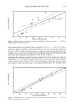
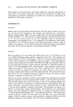
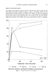
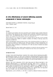
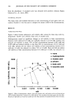
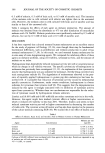
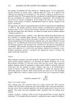
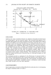
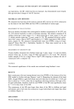
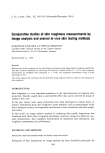
IN VITRO EFFECT OF WHITENING COSMETICS 365 about 54% at 5.0 mM. Ascorbic acid did not markedly reduce cell viability at final concentrations between 0.01 mM and 0.5 mM, but reduced it to about 78% at 1.0 mM and to about 35% at 5.0 mM. TYROSINASE ACTIVITY PER WELL Figure 3 shows human melanocyte tyrosinase activity after culture for three days with hydroquinone, linoleic acid, arbutin, kojic acid, or ascorbic acid. The activity was calculated within concentration ranges that did not markedly reduce cell viability. Hydroquinone dose-dependently reduced tyrosinase activity per well at final concentra- tions between 0.00 ! mM and 0.0 ! mM. Linoleic acid did not reduce tyrosinase activity at final concentrations between 0.001 mM and 0.005 mM. Arbutin dose-dependently reduced tyrosinase activity at final concentrations between 0.01 mM and 1.0 mM. Kojic acid and ascorbic acid showed similar dose-inhibitory curves they did not markedly reduce tyrosinase activity at final concentrations between 0.01 mM and 0.50 mM, but rapidly reduced it at higher concentrations. CELLULAR MELANIN CONTENT Cell pellets were obtained after human melanocytes were cultured for three days with o none * hydroquinone x linolei½ acid -• 3 -e-arbutin • kojic acid • • ascorbic acid _ 2 ........ I . ß . . ....I ...... ill . • ß • 0.001 0.010 0.100 1.000 rnM Figure 3. Effects of hydroquinone, arbutin, kojic acid, ascorbic acid, and linoleic acid on tyrosinase activity per well in human melanocytes. Cultures of 12,500 cells/era 2 were incubated with these agents for three days. Results are expressed as units of mushroom tyrosinase per well. Bars represent of standard deviation from the mean. z o • 0

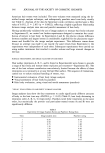
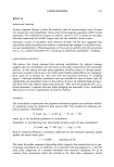
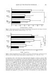
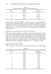
366 JOURNAL OF THE SOCIETY OF COSMETIC CHEMISTS 0.5 mM of arbutin, 0.5 mM of kojic acid, or 0.5 mM of ascorbic acid. The intensity of the melanin color in cells cultured with arbutin was lighter than in the untreated cells. However, the melanin color in cells cultured with kojic acid or ascorbic acid was similar to that of the untreated cells. Table I compares the effects of each agent on melanin production. The amount of melanin was obtained from the absorbance at 475 nm after dissolution of intracellular melanin with 1N NaOH. Melanin production was significantly reduced by 0.5 mM of arbutin, but not by 0.5 mM of kojic acid or 0.5 mM of ascorbic acid. DISCUSSION It has been reported that cultured neonatal human melanocytes are an excellent source for the study of pigment cell biology (15,16), even though there may be fundamental biochemical differences, such as proliferation and melanin production in adult versus neonatal melanocytes (17,18). In this study, we used neonatal human melanocytes for in vitro assay of some depigmenting agents. We compared the inhibitory effects of these agents on melanin synthesis, using cell viability, tyrosinase activity, and the amount of melanin as an index. Hydroquinone dose-dependently reduced tyrosinase activity per well at concentrations at which no change in cell viability was seen. The specific cytotoxicity of hydroquinone in melanoma has previously been investigated (11, 12). An explanation of this cytotoxicity may be that hydroquinone acts as a substrate for tyrosinase, thereby being converted into toxic semiquinone radicals (9). The degradation of melanosomes observed in the pres- ence of topically applied hydroquinone in guinea pig skin melanocytes has been de- scribed (10). It is possible that reduction of tyrosinase activity occurs by melanosomal specific cytotoxicity. Furthermore, under certain conditions, hydroquinone is a better substrate for tyrosinase than tyrosine itself (8). This suggests that the depigmentation induced by this agent is strongly associated with its inhibition of tyrosinase activity apart from cytotoxicity. Whether these two mechanisms are responsible for the reduc- tion of tyrosinase caused by hydroquinone remains unknown. Linoleic acid (0.001 mM and 0.005 mM) did not reduce tyrosinase activity in the wells. However, this agent might have marked cytotoxic effects at higher concentrations, at which it reduced cell viability to less than 20%. Therefore, linoleic acid seems to have reduced tyrosinase activity per well at higher concentrations by decreasing the number of viable cells. The cytotoxic action of linoleic acid has also been reported in the presence of the culture medium containing EGF (19). We studied the in vivo effect of linoleic acid Table I Effects of Arbutin, Kojic Acid, and Ascorbic Acid on Melanin Content in Human Melanocytes Melanin per cell Agent Concentration (ng/cell) Control 0.794 + 0.032 Arbutin 0.5 mM 0.592 + 0.012' Kojic acid 0.5 mM 0.768 + 0.162 Ascorbic acid 0.5 mM 0.746 -+ 0.004 Cultures of 40,000 cells/well were incubated with these agents for three days. Results are expressed as ng of melanin per cell and represent mean -+ SD. * P 0.001, vs control.
Purchased for the exclusive use of nofirst nolast (unknown)
From: SCC Media Library & Resource Center (library.scconline.org)