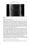
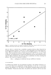
SURFACTANT-SKIN INTERACTIONS 305 porcine skins, and the fluorescence of ANS and tryptophan were measured using the ISS-K2 multifrequency cross-correlation phase and modulation fluorometer (ISS Inc., Champaign, Illinois) with a 300-watt Xenon light source. The corneum or the skin was then treated with the test solutions. The changes in the measured fluorescence properties were related to the corneum protein binding ability of the test product. The experi- mental details are as follows. Stratum corneum. A rectangular piece (2 cm x 1 cm) of stratum corneum was clamped between nylon mesh and immersed in an aqueous solution of ANS (1.6 x 10- 4 moles/l) for one hour at the experimental temperature. This was followed by a thirty-second rinse in distilled water with agitation. The mesh was then removed, and the stratum corneum was placed on spectra mesh to dry under atmospheric condition. The treatment protocol involved clamping the corneum between nylon meshes and immersing them in test solutions at the experimental temperature for a given length of time. After treatment, the corneum was rinsed in distilled water with agitation for thirty seconds. The mesh was then removed, and the corneum was placed on spectra mesh to dry under atmospheric humidity. Corneum soaked in water at the experimental conditions was used as the control. Steady-state fluorescence experiments with isolated corneum were performed in the front surface mode in a blackened triangular cuvette holder. Stratum corneum was placed between the black aluminum surface and a glass slide. The illuminated surface was oriented about 30 ø to the incident beam. The fluorescence was observed at a 90 ø angle from the exciting light. The contribution from scattered light was eliminated by using a cross-polarization condition (vertically polarized excitation with the emission polarizer oriented in a horizontal position) and an appropriate emission high pass cut-off filter. The glass slide attenuated the emission but otherwise did not change the spectral characteristics. As we were interested in the relative change in the emission before and after treatment and not in the absolute magnitude of the intensity, the presence of the glass slide did not affect the results. The excitation and emission monochrometer slits were set at 16 and 4 nm, respectively. The excitation wavelength for tryptophan was at 295 nm, and the corresponding emission was monitored in the 320 to 400 nm region. For ANS, the excitation was done at 370 nm, and the emission spectra were monitored in the 400-550 nm region. Dermatomed skin. Dermatomed porcine skin was cut into 1-cm-diameter discs and placed in a water vapor transmission cell (TEWL) adapted from that described by Blank et al. (16), with a nylon screen underneath the skin for support. The top (epidermal) surface of the skin was sprayed with 200 !xl of an ANS in ethanol solution (5.3 x 10⁻⁴ moles/l) and then washed with 1 ml of distilled water at 37øC. The epidermal side of the skin was then contacted with 1% production solution for one minute, followed by a one-minute rinse with distilled water. The treatment and the rinse were carried out at 37øC. Steady-state fluorescence. Steady-state ANS fluorescence from dermatomed skin was mea- sured with a bifurcated quartz fiber optics probe (C Technologies, Verona, NJ) bundle connected to the ISS K2 fluorometer. The fiber optic bundles in the inner core were used to transmit the excitation light, and the fibers in the surrounding outer core were connected to an emission monochrometer. The open end of the fiber optics bundle was placed vertically on the top surface of the skin. To overcome the problem of total

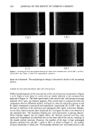
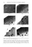
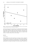
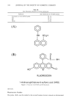
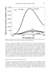
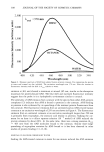
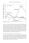
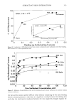
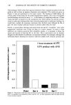
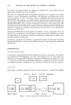
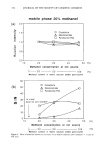
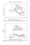
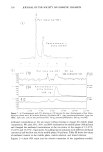
306 JOURNAL OF THE SOCIETY OF COSMETIC CHEMISTS internal reflection of the light at the dermal side, water was used as the optical medium on the dermal side of the skin. Surfactant Adsorption The procedure will be described in more detail in a separate communication (17). Briefly, three 8-mm discs of human stratum corneum were placed between two 1-inch- square teflon screens. These screens were then placed in treatment solutions containing a combination of •4C (specific activity 1 IxCurie/ml) labeled and nonlabeled surfactants. The concentrations of labeled surfactants in the treatment solutions were 3.47 X 10 -4 moles/l, 3.03 x 10 -4 moles/l, and 1.59 x 10 -4 moles/l, respectively, for SLS, SLI, and TEA-Na laurate. The concentrations of unlabeled fraction were varied to obtain total initial surfactant concentrations in the range of 2 x 10⁻³ moles/l to 1 x 10⁻¹ moles/1. The TEA concentration in Na-laurate solution was maintained at 4 x 10⁻² moles/l to maintain the pH and to avoid the precipitation of the Na-laurate. Before the addition of the stratum corneum sample to the treatment solution, 5 Ixl of the treatment solution was removed to provide the initial concentration. After one hour, the screens were rinsed by moving them back and forth in a dish of distilled water five times. Excess water was removed by blotting with a paper towel, and the screens were then allowed to dry overnight in a desiccator. The weight of each individual corneum sample was recorded using a Perkin-Elmer AD4 autobalance, and the sample was placed into scintillation vials containing 0.5 ml distilled water. Scintiverse BD (Fisher Scientific) was added to each vial, and the radioactivity of the treatment solution before and after inclusion of the corneum was determined using liquid scintillation (Beckman). The amount of surfactant bound was calculated and normalized to the final dry weight of the corneum. This procedure corrects for extraction of soluble materials by the surfactant solution, which can be significant for damaging surfactants (17). RESULTS ANS FLUORESCENCE IN SOLUTION The probe ANS has very weak fluorescence (quantum yield 0.004) in water, with a broad emission maximum at about 520 nm. In a less polar solvent, e.g., in octanol, ANS fluoresces much more intensely (quantum yield 0.646), with an emission maxi- mum of 464 nm. The fluorescence intensity also increases significantly when ANS is bound to certain proteins such as apomyoglobin or apohemoglobin (11) or tubulin (18), with the emission maxima shifting to shorter (450-490 nm) wavelengths. The absorp- tion maximum for protein-bound ANS is around 360 nm (11). The fluorescence en- hancement is believed to be due to binding of ANS to specific binding sites in proteins. The spectra of ANS equilibrated in 1% solutions of several pure surfactants, along with the corresponding one in water, are shown in Figure 2. These surfactant concentrations are well above their cmc values (SLS 0.23%, SLI 0.13%, Ci2EO 6 0.003%). The concentration of ANS in these tests was only 1.47 X 10 -4 M (0.004%). Since the ANS concentration is two to three orders of magnitude lower than the miceliar concentration, the solubilization or the lack of it of ANS in these miceliar solutions is dependent only on the affinity of ANS for the micelies. As expected, ANS exhibits low fluorescence
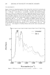
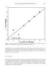
Purchased for the exclusive use of nofirst nolast (unknown)
From: SCC Media Library & Resource Center (library.scconline.org)