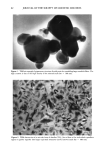
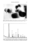
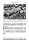
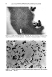
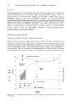
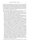
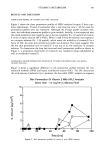
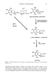
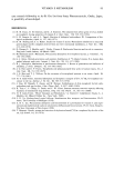
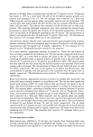
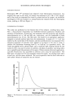
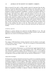
68 JOURNAL OF THE SOCIETY OF COSMETIC CHEMISTS Figure 11. At higher magnification (TEM), the platelet-like TiO 2 crystals of sunscreen preparation 2 seem unagglomerated (circles), even after application to the skin (Co = corneocyte) (scale bar = 100 nm). Figure 12. In sunblock preparation 3, the mineral ingredient appears widespread in the sun care product (TEM) (scale bar = 100 nm).

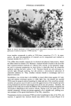
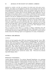
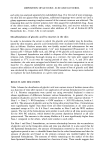
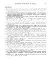
PHYSICAL SUNSCREENS 69 Figure 13. Spatial distribution of TiO 2 particles around desquamated corneocytes (Co), after topical application of sunblock preparation 3 (TEM) (scale bar = 1 }xm). tissue interface corresponds in reality to TiO2/tissue interactions (15-17). In conse- quence, the good biocompatibility of titanium can be extrapolated and widened to titanium dioxide (10,11, 15-17). TiO 2 differs from metallic titanium by its chemical and physical characteristics, which are rather related to ceramics (15). Moreover, titanium dioxide is known to be one of the most corrosion-resistant materials (17). Minute TiO2 crystals, in the nanometer range, are used in sunscreen preparations for their ability to physically block the ultraviolet radiation reaching the skin's surface (7, 18). Titanium dioxide not only attenuates ul- traviolet light through scattering, but also presents a semiconductor absorption of ultraviolet radiation (5). Thus, ultrafine titanium dioxide is increasingly used as a physical sunscreen. Nevertheless, two criteria have to be fulfilled to ensure effectiveness against UV radi- ation and produce minimum opacity to visible light: first, the mineral raw material (TiO2) must be properly selected regarding the shape and size of the crystallites second, extensive precautions must be taken to avoid the agglomeration of the ultrafine particles during formulation and after topical application of the sun care products. Therefore, the purpose of this study was to assess the different TiO2 distribution patterns of applied sun care products depending on the cosmetic vehicles. Owing to the very small sizes of the mineral compound, electron microscopy appeared to be a valuable tool, yielding accu- rate estimates of the spatial distribution of the TiO• crystallites. Transmission electron microscopy (TEM) provided morphological data of the TiO2 raw material. It appeared that titanium dioxide may exhibit either acicular (Figure 2) or
Purchased for the exclusive use of nofirst nolast (unknown)
From: SCC Media Library & Resource Center (library.scconline.org)