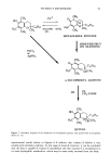
J. Soc. Cosmet. Chem., 47, 85-95 (March/April 1996) Metabolism of vitamin E during skin permeation AE-RI CHO LEE and KAKUJI TOJO, Controlled Drug Delivery Research Center, College of Pharmacy, Rutgers University, Piscataway, NJ 08855-0789. Received November 1995. Synopsis Metabolism of vitamin E (o•-tocopherol) in hairless mouse skin was investigated. An in vitro skin permeation study was performed with [ •H] 2-1,3-o•-tocopherol. Hairless mouse skin was mounted between the half cells of the Valia-Chien diffusion cell, and the total radioactivity in the receptor solution as a function of time was identified with HPLC in line with a UV spectrometer and a liquid scintillation counter. Significant amounts of radioactivity were recovered as o•-tocopherol quinone and more hydrophilic metabolites. To confirm the metabolism of vitamin E in the skin, a stability study of vitamin E in the receptor solution without skin was performed, and most of the radioactivity was recovered as o•-tocopherol, suggesting that major metabolism takes place in the skin, not in the receptor solution. The present study suggests that vitamin E undergoes very extensive metabolism during the skin permeation process and that the skin possesses the enzymes responsible for vitamin E metabolism like other tissues. INTRODUCTION Since its isolation from wheat germ oil in 1920 (1), vitamin E has been the subject of extensive research and considered an important lipid-soluble, membrane-bound antiox- idant. It is known to stabilize cellular membranes by preventing lipid peroxidation (2,3). Vitamin E has also been used in many skin care products claiming anti-aging and moisturizing effects (4). Most skin penetration studies have been conducted to demon- strate clinical effects after dermal application of vitamin E, but no quantitative study of skin penetration of vitamin E has been reported. In 1968, Kamimura et al. investigated the histological distribution of [•4C]-ot-tocopheryl acetate and reported that ot-tocoph- eryl acetate was well absorbed into the stratum corneum and into all the layers of the epidermis and dermis with high affinity around blood vessels (5). Kleck also reported that the percutaneous absorption of 5 % vitamin E in ethanol solution showed that 0.2 % of an applied dose was detected in chamber liquid after 16 hours (4). Klein Ae-Ri Cho Lee's present address is College of Pharmacy, Duksung Women's University, Ssangmun-dong, Dobong-ku, Seoul 132-714, Korea. Kakuji Tojo's present address is Kyushu Institute of Technology, College of Computer Science and Systems Engineering, Iizuka, Fukuoka 820, Japan. 85

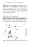
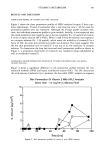
86 JOURNAL OF THE SOCIETY OF COSMETIC CHEMISTS studied the dermal penetration and systemic distribution of [•4C]-labeled vitamin E and reported that vitamin E penetrated the skin very well and that a high accumulation of radioactivity in the skin was observed (6). Until now, skin penetration studies of vitamin E have been performed with radiolabeled vitamin E, and the total radioactivity through the skin has been assayed. Since a major route of skin penetration is through lipid-rich intercellular routes, drugs that have good lipid solubility and favorable oc- tanol/water partition coefficients transfer into the skin readily (7). Based on the above studies, it has been generally concluded that vitamin E penetrates the skin very well. However, we observed that very lipophilic vitamin E (P. C. octanol/water = 476) did not appear in the receptor solution after quite a long lag time and showed a very small permeation rate in HPLC analysis. The significant difference in the penetration profile was observed between HPLC assay and radiotracer analysis. The radiolabeled vitamin E promptly penetrated the skin, while the nonlabeled compound appeared after a remark- able long time lag (about 24 hours). Although there is some evidence for the existence of vitamin E metabolites such as tocopherol quinone, tocopherol hydroquinone, dimers, and trimers in other tissues (8-10), no metabolism study for vitamin E in the skin has been reported. To investigate the possible explanation for these observation, the metabolism of vitamin E in hairless mouse skin has been investigated. MATERIALS AND METHODS Vitamin E (o•-tocopherol), o•-tocopherol quinone, and radiolabeled vitamin E (•[H] 2- dl-tx-tocopherol, 2.0 mCi/mM) were kindly provided by Hoffmann-LaRoche (Nutley, NJ). The purity of radiolabeled compounds was tested by TLC or HPLC in line with a liquid scintillation counter (Rack Beta 1214-001, LKB Instruments Inc., Gaithersburg, MD). Tween-80 (ICI Americas, Inc., Wilmington, DE) and silicone fluid (Dow Corn- ing 360, 20 cp) were used as obtained. All other chemicals were reagent grade. The solvent used in the HPLC assay was HPLC grade. Water was purified by a Nanopure water purification system (Sybron/Barnstead, Boston, MA). Bioflour (E.I. Dupont NEN Research, MA) was used as a liquid scintillation cocktail. ANALYTICAL METHODS HPLC analysis. When the mobile phase of methanol:chloroform (90:10) was used for vitamin E analysis, the vitamin E peak was very sharp at a retention time of about two minutes. To separate vitamin E and its metabolites, we increased the water fraction of the mobile phase to prolong the retention time, resulting in better separation of vitamin E from its metabolites. Methanol:water (95:5) was selected as a mobile phase. The retention time of vitamin E under this condition was about 12 minutes. The assay conditions are summarized in Table I. Radioactivity counting. A liquid scintillation counter (LSC) was used to quantify the concentration of radiolabeled vitamin E. For the assay, the samples (30-80 Ixl) were withdrawn and mixed well with Bioflour scintillation cocktail (5-10 ml). The amount of radioactivity (disintegration per minute, dpm) was determined, and the correction for quenching was made automatically by comparison against a standard quench curve.
Purchased for the exclusive use of nofirst nolast (unknown)
From: SCC Media Library & Resource Center (library.scconline.org)