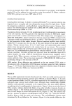
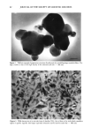
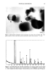
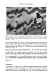
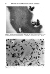
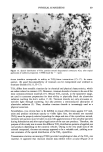
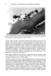
PHYSICAL SUNSCREENS 71 (up to 50m2/g -•) of the ultrafine TiO2, the crystallites remained nonagglomerated and loosely packed in the microspheres (Figure 8). Although the various titanium dioxide raw materials (acicular or prismatic) presented different spatial arrangements within the oil droplets, the TiO 2 crystals appeared to be mainly encapsulated in those round bodies (H•liosides©), thus isolated from the aqueous matrix. After topical application to skin, the oil droplets, containing the physical sunscreen, may burst and produce a quite homogeneous distribution of the TiO 2 crystallites along the stratum corneum (Figures 7,9-11). No particular microstructure was evident in sunblock preparation 3. The titanium dioxide crystals occupied the whole cream layer (Figure 12). The mineral ingredient seemed nonagglomerated and widespread. After application of sunblock preparation 3 onto the skin, either thick (Figure 13) or thin (Figure 14) crystalline coatings of the outer horny layer could be noticed. In spite of the different crystal arrangements (Figures 6, 8) within the oil droplets (H61iosides©), both sunscreen preparations 1 and 2 present similar spatial distributions of the TiO 2 after application to skin, that is a thin and almost monocrystalline coating of the stratum corneum (Figures 7, 10). These thin oxide layers seem suitable because of the organic filter/mineral screen synergy offered by those formulations. On the other hand, sunscreen preparation 3, prepared solely with physical sunscreen agents without any chemical sunscreen added, generally showed the inorganic powder randomly dis- tributed within a thicker layer (Figures 12, 13) compared to sunscreen preparations 1 and 2. Furthermore, it must be pointed out that the ultrafine titanium dioxide crys- tallites appeared to be exclusively confined to the outer surface of the stratum corneum and around desquamating corneocytes (Figures 10, 13). In summary, our observations provide conclusive evidence that the spatial distribution of a physical sunscreen is greatly influenced by the formulation of the sun care product. Thus, the sunscreening efficacy is directly related to both the mineral ingredient and the formulation. Moreover, this study emphasizes the contribution of electron microscopy in determining the location and behavior of TiO 2 crystals upon the skin's surface. Such electron-microscopic assessments yield useful information for quality checks of physical sunscreens, thus allowing us to explore product effectiveness. REFERENCES (1) S.-Y. Lin and R.-C. Liang, The effect of ultraviolet B irradiation on the isolated porcine stratum corneum: Colorimetric and ATR/FT-IR spectroscopic investigations, Biotaed. Res., 15 (1) 9-15 (1994). (2) L. A. Applegate and E. Frenk, Cellular defence mechanisms of the skin against oxidant stress and in particular UVA radiation, Eur. J. Dermatol., 5, 97-103 (1995). (3) M. F. Naylor, A L Boyd, D. W. Smith, G. S. Cameron, D. Hubbard, and K. H. Neldner, High sun protection factor sunscreens in the suppression of actinic neoplasia, Arch. Dermatol., 131, 170-175 (1995). (4) R. Marks, P. A. Foley, D. Jolley, K. R. Knight, J. Harrison, and S. C. Thompson, The effect of regular sunscreen use on vitamin D levels in an Australian population, Arch. Dermatol., 131, 415-421 (1995). (5) R. M. Sayre, N. Kollias, R. L. Roberts, A. Baqer, and I. Sadiq, Physical sunscreens, J. Soc. Cosmet. Chem., 41, 103-109 (1990).

72 JOURNAL OF THE SOCIETY OF COSMETIC CHEMISTS (6) M. L. Schlossman, Treated pigments: New ways to impart color on the skin, Cosmet. Toiletr., 105, 53-64 (1990). (7) G. H. Dahms, Formulating with a physical sun block, Cosmet. Toiletr., 107, 87-92 (1992). (8) P. Msika, F. Boyer-Denayrou, and H. Coutelle, Photoprotective microdispersion: A new concept in sunscreen formulation, 18th International IFSCC Congress, Venice, Italy, P 046, 390-401 (October 1994). (9) COLIPA: Sun Protection Factor Test Method ref. 94/289, The European Cosmetic, Toiletry and Perfumery Association (October 1994). (10) S. B. Goodman, J. A. Davidson, and V. L. Fornasier, Histological reaction to titanium alloy and hydroxyapatite particles in the rabbit tibia, Biomaterials, 14, 723-728 (1993). (11) S. Torgersen, G. Moe, and R. Jonsson, Immunocompetent cells adjacent to stainless steel and titanium miniplates and screws, Eur. J. Oral Sci., 103, 46-54 (1995). (12) L. Linder, T. Albrektsson, P-I. Brinemark, H-A. Hansson, B. Ivarsson, U. J/Snsson, and I. Lun- str/Sm, Electron microscopic analysis of the bone-titanium interface, Acta Orthop. Scand., 54, 45-52 (1983). (13) M. A. Listgarten, D. Buser, S. G. Steinemann, K. Donath, N. P. Lang, and H. P. Weber, Light and transmission electron microscopy of the intact interfaces between non-submerged titanium-coated epoxy resin implants and bone or gingiva, J. Dent. Res., 71 (2), 364-371, (1992). (14) E. A. B. Effah, P. D. Bianco, and P. Ducheyne, Crystal structure of the surface oxide layer on titanium and its changes arising from immersion, J. Biom. Mater. Res., 29, 73-80 (1995). (15) J.E. Ellingsen, A study on the mechanism of protein adsorption to TiO 2, Biomaterials, 12, 593-596 (1991). (16) T. Hanawa, "Titanium and Its Oxide Film: A Substrate for Formation of Apatite," in The Bone- Biomaterial Interface, J. E. Davies, Ed. (University of Toronto Press, Toronto, 1991), pp. 51-58. (17) T. Miyazaki, Surface treatments of titanium for a biomedical use, Bull. Kanagawa Dent. Col., 22 (2), 113-123 (1994). (18) J. O. Funk, S. H. Dromgoole, and H. I. Maibach, Sunscreen intolerance: Contact sensitization, photocontact sensitization, and irritancy of sunscreen agents, Dermatologic Clinics, 13 (2) 473-480 (1995). (19) H. Schatz, A.M. Kligman, S. Manning, and T. Stoudemayer, Quantification of dry (xerotic) skin by image analysis of scales removed by adhesive discs (D-Squames), J. Soc. Cosmet. Chem., 44, 53-63 (1993).
Purchased for the exclusive use of nofirst nolast (unknown)
From: SCC Media Library & Resource Center (library.scconline.org)