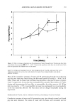
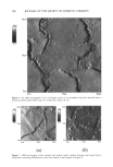
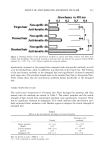
ABSORPTION OF AHAs IN SKIN 189 Table I Composition of the Oil-in-Water Emulsions for 5% Alpha Hydroxy Acids Formulation A Grams per 100 grams emulsion Phase A Polyoxyethylene (100) glycerol stearate (ICI Surfactants, Wilmington, DE) 2.0 Mineral oil (light) (Penreco, Karns City, PA) 10.0 Cetearyl alcohol (Henkel Corp., Hoboken, NJ) 3.0 Phase B Laureth-4 (Lipo Chemicals, Paterson, NJ) 1.0 Propylene glycol (Aldrich Chemical Co., Milwaukee, WI) 5.0 Alpha hydroxy acid 5.0 Phthalate-HC1 buffer* Potassium phosphate-NaOH** 73.0 Phase C Preservative Methyl-p-hydroxybenzoate (Pfaltz & Bauer, Inc., Stamford, CT) 0.5 Propyl-p-hydroxybenzoate (Pfaltz & Bauer, Inc., Stamford, CT) 0.5 * Phthalate-HC1 buffer (pH = 3): 50 ml of 0.1 M potassium bi- phthalate + 22.3 ml of 0.1 M HC1 diluted with water to 100 mi. ** Potassium phosphate-NaOH buffer (pH = 7): 50 ml of 0.1 M potassium phosphate + 29.1 ml 0.1 M NaOH diluted with water to 100 mi. appropriate amounts of AHA were added to aliquots of the stock emulsion to give the desired concentration of AHA. PERCUTANEOUS ABSORPTION EXPERIMENTS Skin absorption studies were conducted by using human skin freshly obtained from abdominoplasty procedures. The skin was placed in a saline solution at the clinic and kept in cool packing as it was transported to the laboratory and transferred to Hepes buffered Hanks' balanced salt solution (HHBSS). Subcutaneous fat was removed from the skin, and the surface was gently cleaned with a 10% soap solution and rinsed with distilled water. The skin was mounted on a Styrofoam block and cut with a Padgett dermatome (Padgett Instruments, Dermatome Division, Kansas City, MO) to a thick- ness of 200-340 pm. Skin discs were prepared with a punch and placed epidermis-side up in Teflon flow-through diffusion cells (5). Prior to assembly, the flow-through diffusion cell system was disinfected with 70% ethanol and rinsed with receptor fluid. The diffusion cells were maintained at 35øC in an aluminum holding block heated by a circulating water bath this maintained the surface temperature of the stratum corneum at 32øC. The skin was perfused with HHBSS, pH 7.4, receptor fluid at a flow rate of 1.5 ml/h to maintain the viability of the skin in the diffusion cells for the duration of the 24-h study (6).

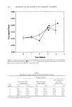
190 JOURNAL OF THE SOCIETY OF COSMETIC CHEMISTS A 20-min skin barrier integrity check, using [3H]water, was conducted prior to the application of the AHA test formulations to ensure that the permeability of the human skin was in the normal range and that the skin was not damaged (7). Cells in which the percent of the applied dose of [3H]water absorbed through the skin was greater than the historical limit of 0.35% were discarded. The AHA test formulations were previously prepared to give an average dose of 0.55 laCi of [•4C] radiolabeled AHA per cell. The emulsion was applied to the skin at 3 mg/cm 2 of exposed skin in the diffusion cells (exposed skin -- 0.64 cm2). At the end of each experiment the skin surface was washed three times with 0.3 ml of a 10% soap solution and rinsed three times with 0.3 ml of distilled water to remove unabsorbed material remaining on the surface of the skin. The skin was removed from the diffusion cell and tape-stripped with Scotch Magic TM cellophane tape (3M Commercial Office Supply Division, St. Paul, MN) ten times to remove the stratum corneum. The remaining epidermis was separated from the dermis with heat. The skin was wrapped in Saran Wrap plastic wrap (DowBrands L.P., Indianapolis, IN) and submerged in a 60øC water bath for 40 s. The skin was unwrapped and the epidermis was then slowly peeled from the dermis. The epidermis and dermis were cut into thin strips with a razor and digested with tissue solubilizer. ALPHA HYDROXY ACID ANALYSIS The absorbed radioactivity in the 6-h receptor fluid fractions and the skin layers was measured by liquid scintillation counting (Minaxi• Tri-Carb © 4000 Series liquid scin- tillation counter, Packard Instrument Co., Downers Grove, IL) using Ultima Gold TM (Packard Instrument Co., Meriden, CT) liquid scintillation cocktail. BARRIER INTEGRITY DETERMINATIONS The barrier integrity of hairless guinea pig skin following 24-h exposure to glycolic acid formulations was assessed by measuring the steady-state rate of penetration of [•H]water and then calculating a permeability constant (Kp). Skin from 4- to 6-month-old male hairless guinea pigs [strain Crl:AF/HA (hr/hr)Br] (Charles River Laboratories, Wilming- ton, MA) was dermatomed to a thickness of 200-300 pm and assembled into flow- through diffusion cells. Glycolic acid formulations were applied to the surface of the skin (3 mg/cm2), while some diffusion cells containing skin were left untreated (control skin). After 24 h, the surface of the skin (including untreated control skin) was washed three times with 0.3 ml of a 10% soap solution, rinsed three times with distilled water, and blotted dry with a cotton-tipped applicator. [•H]water (2.34 to 2.88 pCi) was applied in excess (800 pl) to the surface of the skin, the diffusion cell was covered, and effluent from the flow cell was collected every half hour until a steady-state rate of permeation was established (about 4 to 4.5 h). Permeability constants were calculated by dividing the rate by the initial concentration of [•H]water. SKIN SURFACE pH MEASUREMENTS The pH profile of human skin in flow-through diffusion cells was determined 24 h after
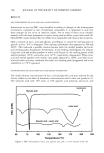
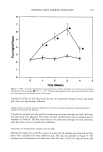
Purchased for the exclusive use of nofirst nolast (unknown)
From: SCC Media Library & Resource Center (library.scconline.org)