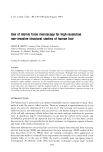
200 JOURNAL OF THE SOCIETY OF COSMETIC CHEMISTS conditions for successful imaging, which could damage or alter the appearance of fine cuticular structures. Atomic force microscopy (AFM) is a relatively new technique (10) and is a member of a generic class of related scanning probe microscopy (SPM) techniques (11,12). The principle of AFM relies on the use of a sharp, pyramidal tip mounted on a cantilever that is brought into close proximity to the sample of interest where intermolecular forces acting between the tip and the surface cause the cantilever to bend. Cantilever deflec- tions are detected by a laser beam focused on the top of the cantilever and are converted to a digital voltage signal that is then applied across a piezoelectric ceramic element to which the sample is mounted. This has the effect of maintaining a constant force, in the order of a few nano-Newtons, between the scanning tip and the sample. A three- dimensional map of the specimen's topography can therefore be obtained by plotting this feedback voltage signal against X and Y coordinates. Confocal microscopy has recently been demonstrated to be a good technique for imaging hair surfaces in their natural environment (13). However, the resolution limit for this method is of the order of 0.25 pm, an improvement on conventional light microscopy (0.6 pm), but much less than that of electron microscopy. Since AFM does not require the focusing of light or other radiation for its operation, a very high resolution is achievable, of the order of 1-2 nm for biological materials. This high resolving power, together with the ability to image surfaces in aqueous environments, makes AFM a very attractive technique for imaging hair surfaces. Only a few reports on the use of AFM in this application area have appeared in the literature (14-17). Here, the potential applications of AFM for structural investigations of human hair surfaces are discussed. MATERIALS AND METHODS Human scalp hairs were obtained from biopsies from European brown-haired subjects who had washed their hair approximately 5 h prior to the investigations. A sodium lauryl sulphate-based shampoo and conditioner containing dimethicones or 1% zinc pyrithione was used, and hair was allowed to dry naturally at room temperature. 'Un- washed' samples refer to hair that had been washed the previous day and had not been groomed. Hair sections of 1 cm in length, no further than 5 cm from the scalp, unless otherwise stated, were cut and fixed on a sample mounting assembly using carbon- loaded double-sided adhesive tape. AFM studies were performed using a Discoverer TMX2000 scanning probe microscope (SPM) (TopoMetrix Corporation, Saffron Wal- den, Essex, UK) under atmospheric conditions, although in some experiments, a liquid cell was used to obtain images in an aqueous environment. A relative humidity of 60-65% was noted during all the experiments, although this was not accurately con- trolled room temperature was recorded to be 22øC. Standard profile pyramidal silicon nitride tips mounted on cantilevers of force constants in the range 0.036 to 0.072 N m -• were used. The sample was held in position on a piezoelectric tripod scanner capable of a maximum XYZ translation of 75 x 75 x 12 pm. Images were left-shaded to enhance topographical features and displayed on a monitor with a resolution of 500 lines x 500 pixels. Quantitative data, such as height measurements and surface roughness, were obtained using TopoMetrix image analysis software (18). The arithmetic surface rough-

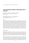
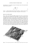
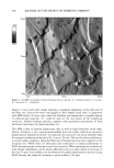
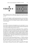
AFM FOR STUDIES OF HUMAN HAIR 201 ness average, Ra, is the arithmetic average of the absolute values of the measured profile height deviations, given by: n •lZ i - Zl i=l Ra = (1) n where, n -- number of height positions along line profile, Z i -- height at position i (nm), -- and Z -- average height (nm). For this study, a line profile of 4 pm was used. RESULTS AND DISCUSSION Figure 1 shows a three-dimensional AFM micrograph of the surface of a typical human scalp hair taken from a European brown-haired subject. Cuticles were found to be very well defined and lying flat on the hair shaft. The arrangement of cuticular cells is multilayered, due to cuticles overlapping and not because of multiple cell layering. An interesting scratch mark feature can be observed traversing from the top left to the bottom right in Figure 1 in the general direction of the fiber axis such features have been observed elsewhere (19). On closer examination of the image, three types of cuticular cell can be identified: the exocuticle, the endocuticle, and the marginal band (A- or u-layer). These structures are indicated in Figure 2. The marginal band (20,21), denoted "A" covered a large part of the exocuticle and was very smooth in appearance. The thickness of this layer was typically in the range of 30-70 nm, in agreement with the 45-50 nm range of values reported elsewhere (22). The exocuticles, denoted "B" in ..-: • [ 26,52 23 nm 1326.12 nm 0 nm . '.." i :'-._ ,,. ß .. ß . . 20•m•......... '. : . , •' , . Ol•m Ol•fn Figure 1. Three-dimensional representation of an AFM micrograph of human European brown scalp hair.
Purchased for the exclusive use of nofirst nolast (unknown)
From: SCC Media Library & Resource Center (library.scconline.org)