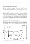
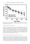
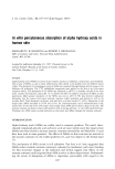
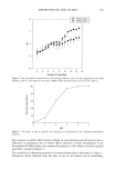
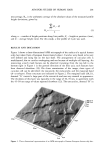
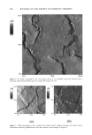
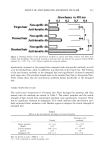
202 JOURNAL OF THE SOCIETY OF COSMETIC CHEMISTS ß -90.0 ø 90.0 ø 20 prn .... ,' ( 10 prn A 0pm , ,: ::-,, 0 prn B B A B B 10 pm 20 pm Figure 2. An AFM micrograph of human European brown scalp hair. A = marginal band (A- or c•-layer), B = exocuticle, C = endocuticle. Figure 2, were much more rough, showing a woodgrain appearance in the direction of the fiber axis. Exocuticles were very jagged at their broken distal ends, in agreement with SEM studies. In some cases, where the breakage was fragmented, a rougher deposit of endocuticular material, "C," could be seen on the top surface of the underlying exocuticle. Similar breakage patterns, together with postulated mechanisms of their formation, have been discussed elsewhere (3). The AFM is able to provide quantitative data as well as high-resolution visual infor- mation. In Figure 3, two cross-sectional profiles show the surface relief of an exocuticle (upper) and an endocuticle (lower). As expected, the exocuticle was much smoother than the exposed underlying endocuticle (Ra: 9 nm cf. 30 nm). The use of surface profilometry to record surface roughness along the fiber axis of human hairs has been reported in the literature (23). While there are obviously some similarities in surface profilometry to AFM, the macroscopic technique is much less sensitive. When operating on a submicron scale, height information can be grossly affected by the very large radius of curvature of the tip (2.5 Fm). Such artefacts are not observed in profile measurements obtained using AFM, because the radius of curvature is much smaller (50 nm).

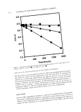
AFM FOR STUDIES OF HUMAN HAIR 203 The unprecedented high resolution of AFM offers the potential of investigating the effects of different hair-care products and treatments on the topography of human hair. For example, when the same hair sample as that shown in Figure 1 was treated with an alternative product containing the antimicrobial agent, 1% zinc pyrithione (24), a fine microgranular deposit was observed on exocuticle surfaces (Figure 4). This deposit was found in other similarly treated hair samples, including those from other subjects, and was very different in appearance to the endocuticular material observed in Figure 1. Such a deposit may reduce the luster of the hair. One of the most important advantages of AFM is its ability to image samples in various environments. The technique can not only image surfaces under atmospheric conditions, unlike SEM and TEM, which require high vacuum, but can also probe surfaces in an aqueous environment. Hair specimens can very easily be imaged in an environmental chamber, or "wet cell", offering the potential for real time, in situ studies of various phenomena such as cleansing, swelling, damage, and repair. Figure 5 shows the same hair sample as that shown in Figure 4, but imaged in distilled water. The image retains the same high resolution as that obtained in air, showing the woodgrain topography of the exocuticles. Almost all of the deposit thought to be zinc pyrithione is absent from the hair surface, although some remains of the endocuticle are still visible. This further supports the fact that the microgranular deposits observed in Figure 4 cannot be ascribed to endocuticular material. The distal ends of the exocuticles were found to be raised with respect to the underlying surface (1.2 pm compared to 0.2 pm in air for the same hair sample) due to swelling.
Purchased for the exclusive use of nofirst nolast (unknown)
From: SCC Media Library & Resource Center (library.scconline.org)