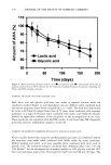
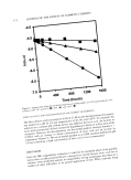
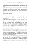
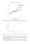
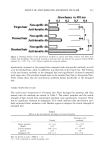
ABSORPTION OF AHAs IN SKIN 197 The percutaneous absorption of GA through animal skin has previously been reported from an aqueous solution (9). Absorption values of 0.7% and 0.9% were reported in 8 h through minipig and hairless mouse skin, respectively, from a pH 3.8 aqueous solu- tion. But an infinite dose applied to skin of over 100 pl per cm 2 makes the values not relevant to "in use" conditions. We have found that AHAs are extensively absorbed into and through human skin from a relevant dose of an O/W emulsion adjusted to pH 3.0. Approximately 27% of the applied dose ofglycolic acid was absorbed in 24 h, and there was no significant difference in total absorption when compared with values obtained from longer-chain AHAs. REFERENCES (1) D. B. Hagan, D. T. Partort, and A. P. Taylor, A study of the structure-activity relationships present in skin active agents, Int. J. Cosmet. Sci., 15, 163-173 (1993). (2) J.j. Leyden, R. M. Lavker, G. Grove, and K. Kaidbey, Alpha hydroxy acids are more than moistur- izers,J. Geriatr. Dermatol., 3 (Suppl A), 33A-37A (1995). (3) R. M. Lavker, K. Kaidbey, and J. j. Leyden, Effects of topical ammonium lactate on cutaneous atrophy from a potent topical corticosteroid, J. Amer. Acad. DermatoL, 26, 535-544 (1992). (4) E.J. Van Scott and R.J. Yu, Alpha hydroxy acids: Procedures for use in clinical practice, Cutis, 43, 222-228 (1989). (5) R.L. Bronaugh and R. F. Stewart, Methods for in vitro percutaneous absorption studies. IV. The flow-through diffusion cdl,J. Pharm. Sci., 74, 64-67 (1985). (6) S. W. Collier, N.M. Sheikh, A. Sakr, J. L. Lichtin, R. F. Stewart, and R. L. Bronaugh, Maintenance of skin viability during in vitro percutaneous absorption/metabolism studies, Toxicvl. AppL PharmacoL, 99, 522-533 (1989). (7) R. L. Bronaugh, R. F. Stewart, and M. Simon, Methods for in vitro percutaneous absorption studies. VI. Use of excised human skin, J. Pharm. Sci., 75, 1094-1097 (1986). (8) H. •hman and A. Vahlquist, In vivo studies concerning a pH gradient in human stratum comeurn and upper epidermis, Acta Derre. Venereol. (Stockh.), 74, 375-379 (1994). (9) M. Goldstein and R. Brucks, Evaluation of glycolic acid permeation through skin, Pharm. Res., 11, S-180 (1994).

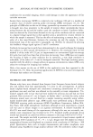
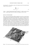
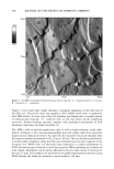
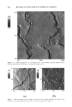
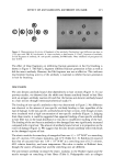
j. Soc. Cosmet. Chem., 48, 199-208 (July/August 1997) Use of atomic force microscopy for high-resolution non-invasive structural studies of human hair JAMES R. SMITH, Scanning Probe Microscopy Laboratory, School of Pharmacy, Biomedical and Physical Sciences, University of Portsmouth, St. Michael's Building, White Swan Road, Portsmouth PO1 2DT, United Kingdom. Accepted for publication September 3 O, 1997. Synopsis The morphology of the fine cuticular structure of human hair has traditionally been investigated using scanning electron microscopy and transmission electron microscopy. Although these techniques are very useful, they require specimens to be coated with metallic films or to be suitably stained. In addition, high vacuum conditions are required that may damage or alter the appearance of delicate cuticular structures. Atomic force microscopy is a relatively new scanning probe technique, capable of imaging surfaces at high resolution under ambient conditions. In this communication, the potential applications of atomic force microscopy for structural investigations of human hair surfaces are discussed. Fine surface structures, such as the exocuticle, the endocuticle, and the marginal band (A- or oMayer), could be easily identified. The technique has also been demonstrated to image hair surfaces in liquid environments, opening the way to in situ studies of the effects of hair-care products and treatments. INTRODUCTION The human hair is protected by an almost formidable barrier comprised of many thou- sands of scale-like plates, called cuticles. These are arranged in approximately six to ten layers (1), each of a thickness of 0.3-0.5 pm (2,3). The cuticular cell consists of essen- tially two layers, the endocuticle and exocuticle (1), the former being the inner one third of the cell and containing cellular debris that can easily undergo hydrolysis by proteases (4,5). A significant degree of keratinization occurs near the surface of the cuticle, and trichohyalin granules (6) are deposited near the lateral cell wall it is this layer, termed the exocuticle, that is responsible for the hair's resistance to detergents, organic solvents, and many other harsh environments (7). Structural information concerning hair cuticles has largely been obtained from scanning electron microscopy (SEM) and transmission electron microscopy (TEM) studies of hu- man hair fibres (3,4,7-9). While these are valuable techniques, their application in structural hair analysis is limited by the requirement for samples to be coated with a thin metallic film, or, in the case of TEM, the need to suitably stain specimens prior to investigation. In addition, both these microscopic techniques require high vacuum 199
Purchased for the exclusive use of nofirst nolast (unknown)
From: SCC Media Library & Resource Center (library.scconline.org)