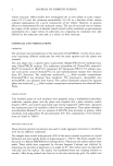
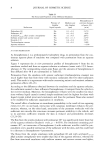
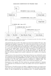
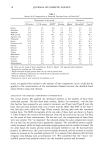
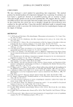
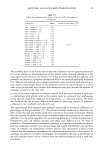
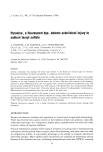
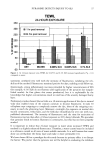
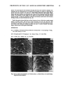
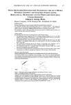
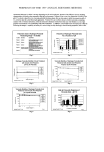
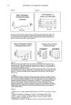
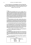
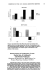
DAMAGED COMPONENTS OF PERMED HAIR 15 Hair CHCI3/MeOH, 16 hours at room temp. I I 1N NaOH/90% MeOH, 2 hours at 60 øC 1% SDS/2M 2-ME, 3 days at 50 øC 1 Integral lipids 1% SDS/0.4M 2-ME, 3 days at 50 øC 1%SDS/0.2M DTT, 6 days at 37 øC r ............................................................[•,, [ [ II-IighmolulaWight i I Cuti'+sidu .. ................................. Cortex ........................................ : Figure 1. Scheme for fractionation of hair components. A 10-mg hair was used in this work, with liquor ratio at 100. See Materials and Methods for details. extracted for three days at 50øC. The residue was removed and washed with water, then lyophilized and weighed. Furthermore, the residue was extracted with a 1-ml solution of 25 mM Tris-HC1 buffer (pH 8.3) that contained 1% SDS and 0.2 M DTT for six days at 37øC to extract the high-molecular-weight protein. After washing and lyophilization, the residue was weighed again. The loss in weight was regarded as the high-molecular- weight protein. The residue included the cuticle. The amounts of the matrix and microfibril protein were determined using the Bio-Rad protein assay (Bio-Rad Lab., Hercules, CA) and compared with a protein calibration curve of BSA in the extraction buffer. Characterization of the extracted fractions. Each extracted fraction was confirmed by amino acid composition and molecular weight. The molecular weight of the extracted protein was determined using Tricine-SDS-PAGE (10). The amino acid analysis was performed according to the Pico-Tag © method supplied by Millipore Co. (Milford, MA). The half-cystine content was estimated as cysteic acid converted by performic acid. Determination of the isopeptide (IP). This method was previously published by Adamski (11). Isopeptide was determined by amino acid analysis after successive peptidase di- gestion. Determination of 18-methyl-eicosanoic acid (MEA). 18-Methyl-eicosanoic acid, which exists only in the cuticle, was analyzed by the method of Wertz (12) with slight modification. Briefly, 10 mg of delipidized hair was hydrolyzed with 1 N sodium hydroxide/90%

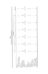
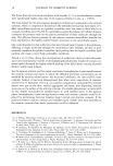
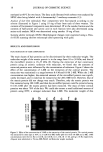
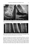
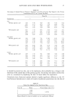
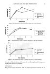
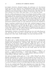
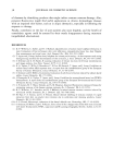
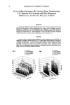
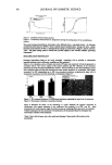
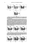
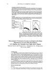
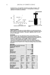
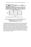
16 JOURNAL OF COSMETIC SCIENCE methanol at 60øC for two hours. The fatty acids liberated with solvent were analyzed by HPLC after being labeled with 4-bromomethyl-7-methoxy-coumarin (13). Analysis of hair from individuals. Hair components were fractionated according to the scheme illustrated in Figure 1 using 10 mg of hair from each of three women. The contents of fractionated components were determined. IP in the residue fraction and the contents of half-cystine and cysteic acid in each fraction were also determined using amino acid analysis. MEA was determined using another 10 mg of hair. Scanning electron microscopy (SEM). Morphological changes were examined using a Hita- chi-S520 scanning electron microscope after sputtering with gold. RESULTS AND DISCUSSION FRACTIONATION OF HAIR COMPONENTS The main classes of hair proteins can be discriminated by their molecular weight. The molecular weight of the matrix protein is in the range from 10 to 30 kDa, and that of the microfibril protein is 45-55 kDa (9). During the extraction of hair constituent protein using an anionic surfactant with reductant, we found that the amounts of extracted proteins were influenced by the concentration of reductant. Figure 2 shows the effect of the concentration of 2-ME on the extraction of the cortical protein. The extraction was maximized in a range between 0.4 M and 0.8 M 2-ME. When the 2-ME concentration was higher, the extracted amount of the microfibril protein was signifi- cantly decreased, and it could not be extracted at the 2M 2-ME level. However, that of the matrix protein did not change very much. Therefore, only the matrix protein was first extracted using 2M 2-ME, and then the microfibril protein could be extracted using 0.4 M 2-ME after the matrix protein had been extracted. The mass of these cortical proteins was about 70% of the hair. We could also extract a small additional amount of protein using DTT, a stronger reductant than 2-ME. The molecular weight of this 97K 67K 43K 31K 22K 14K IlK 6K a b c d e f g Figure 2. Effect of the concentration of 2-ME on the extraction of the cortical protein The cortical protein was extracted for three days at 50øC in 25 mM Tris-HCl buffer (pH 8.3) with 1% SDS and 2-ME. 2-ME concentrations were (b) 0.2 M, (c) 0.4 M, (d) 0.8 M, (e) 1.2 M, (f) 1.5 M, and (g) 2.0 M, respectively. Aliquots were submitted to Tricine-SDS-PAGE. (a) Molecular weight standards.
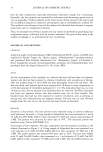
Purchased for the exclusive use of nofirst nolast (unknown)
From: SCC Media Library & Resource Center (library.scconline.org)