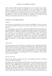
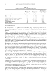
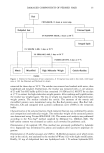
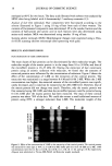
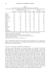
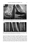
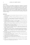
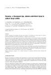
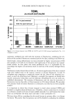
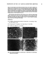
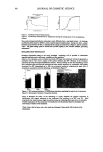
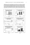
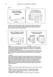
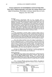
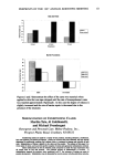
DAMAGED COMPONENTS OF PERMED HAIR 17 protein is more than 100 kDa (Figure 3), and so we called this protein the high-molecular- weight protein (HMW). We confirmed the purity and identity of extracted fractions not only by molecular weight but also by amino acid composition. The amino acid composition of each fraction from the root end hair is summarized in Table I. The extracted protein using 2M 2-ME was in good agreement with the matrix protein, and the 0.4 M extract was the micro- fibril protein when prepared by reduction following alkylation (9). The amino acid composition of HMW, which increased toward the tip end, as discussed later, was similar to that of microfibril protein. The residue was almost the same as the amino acid composition of the cuticle prepared by physical isolation (14,15) in the root end. Figure 3 shows the Tricine-SDS-PAGE of each fraction except the residue. The molecular weight of each fraction was also confirmed, and they showed good purity. When viewed by SEM, the hair from which only matrix protein had been extracted was shriveled but still had a fibrous shape, as seen in Figure 4a. On the other hand, when both the matrix and the microfibril protein were extracted, the hair was no longer fibrous and only the cuticle layers remained just like the sheath (Figure 4b). Conse- quently, we have established a method of fractionating the hair components, as shown in Figure 1. As we have previously described (16), we cotfid not separately extract the matrix and the microfibril protein when the reductant is thioglycolic acid (TGA), and the extracting efficiency is not enough when the surfactant has the properties of low denaturation. We considered that the microfibril protein requires a strong denaturing surfactant for ex- traction since it is a tightly packed structure. Both 2-ME and DTT have alcoholic OH in their molecules, and the denaturing efficiency of the surfactant is weakened if the concentration of these reductants is too high. This is the reason why only the matrix protein was extracted using the concentrated reductant. In fact, the mild surfactants such as sodium dodecyltri (oxyethylene) sulfate cotfid not extract the microfibril protein (16). This is a unique method of isolating the main classes of hair proteins (the matrix and the microfibril). It is an easy, high-yield method that does not need to be modified. In this 97K 67K 43K 31K 22K 14K 11K a b c d 6K Figure 3. Tricine-SDS-PAGE of extracted fractions: (a) molecular weight standards (b) 2 M 2-ME extract (c) 0.4 M 2-ME extract after (b) (d) 0.2 M DTT extract after (c). Refer to Figure 1 for detail.

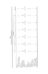
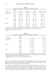
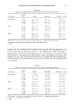
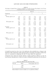
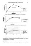
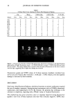
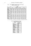
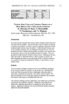
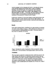
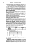
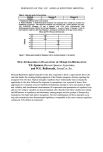
18 JOURNAL OF COSMETIC SCIENCE Table I Amino Acid Compositions of Extracted Fractions From the Root End a Determined in this study Literature 2 M extract 0.4 M extract HMW b Residue Matrix • MicrofibriF Cuticle d Lysine 0.6 3.5 2.0 3.1 0.6 3.5 3.4 Histidine 1.0 0.7 0.5 1.2 0.9 0.7 0.5 Arginine 6.6 5.4 7.0 4.6 5.4 7.1 2.6 Half-cystine e 23.5 9.0 10.1 19.1 27.2 7.6 18.1 Aspartic acid 2.9 8.3 5.9 3.1 2.5 9.3 3.2 Threonine 10.7 6.9 7.9 6.2 10.3 5.4 4.6 Serine 12.0 8.0 11.8 12.6 11.9 8.9 16.1 Glutamic acid 8.4 17.2 12.4 6.4 8.4 16.5 8.9 Proline 12.3 5.1 9.1 7.5 12.7 3.8 10.5 Glycine 6.2 5.2 6.3 11.4 6.1 5.1 8.8 Alanine 2.0 6.3 5.3 4.6 2.3 6.9 5.4 Valine 5.5 6.0 7.0 6.7 5.2 6.1 7.3 Methionine 0.0 1.1 0.2 0.9 0.0 0.4 0.5 Isoleucine 2.0 3.6 3.4 3.0 1.8 3.6 2.2 Leucine 3.3 9.2 7.1 5.5 2.2 10.2 4.5 Tyrosine 1.6 2.5 2.7 2.4 1.5 2.5 2.1 Phenylalanine 1.4 2.0 1.8 1.9 1.1 1.9 1.2 All values are the mean of three experiments. Refer to Figure 1 for experimental conditions. Expressed as residues per 100 residues. High-molecular-weight protein from the tip end of perreed hair. Reduction following alkylation and isoelectrical precipitation method (9). d Physical isolation method (15). Estimated as cysteic acid converted by performic acid. study, we applied this method to the analysis of hair components, but it could also be applied to the reconstitution of the intermediate filament because the disulfide bond easily reforms using only dialysis. ANALYSIS OF THE DAMAGED COMPONENTS OF PERMED HAIR The cortical proteins. We applied the developed method to the analysis of hairs from individual persons. The hair from three women, labeled "no treatment," was the hair that had not been exposed to any cosmetic treatment, and Type I and Type II were the hairs that had been permed every two or three months. More than 80% of the Type I hair had split ends, while the Type II hair scarcely had any split ends but contained broken hairs. The SEM micrographs of the tip ends of these hairs are shown in Figure 5. Table II shows the contents of each fraction, from the root end to the tip end. The data are the mean of three examinations. On the root end, the compositions of three hairs were very similar. The "no treatment" hair showed almost the same composition toward the tip end, but the perreed hairs apparently changed. The microfibril protein signifi- cantly decreased, while the high-molecular-weight protein and the residue increased. On the other hand, the matrix protein slowly decreased whether or not the hair had been permed. As shown later, the cuticle layers actually decreased, and the increase in residue means an increase in the insoluble proteins (9). It is reported that alkaline pH will form irregular cross-linking such as lanthionine (17). We considered that the "intact" mi- crofibril protein had partially turned into the insoluble protein and decreased.
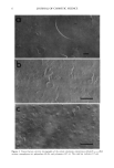
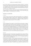
Purchased for the exclusive use of nofirst nolast (unknown)
From: SCC Media Library & Resource Center (library.scconline.org)