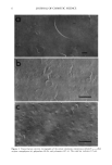
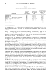
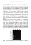
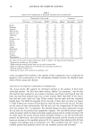
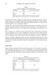
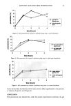
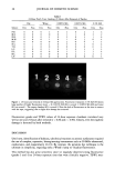
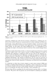
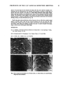
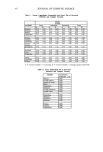
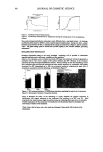
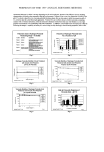
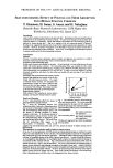
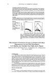
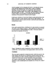
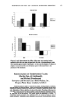
PYRANINE DETECTS INJURY TO SLS 37 350 300 250 200 150 lOO 5O -5O TEWL 24-HOUR EXPOSURE - [] 1 hr post removal _ []24 hrs post removal * ! IIIIIII1.._., .... , , , , DRY WATER 0.005% SLS 0.025% SLS 0.1% SLS Figure 2. At 24-hour exposure sites TEWL for 0.025% and 0.1% SLS increased significantly (*p 0.1) compared to water. surements correlated very well with the intensity of fluorescence, validating the reli- ability of the method. Fluorescence remained positive for days, enabling serial follow-up. Interestingly, strong inflammatory reactions provoked by higher concentrations of SLS (for example, 0.5 % SLS) do not fluoresce after application of the pyranine dye (unpub- lished results). At first glance this seems paradoxical, but it is explainable by the knowledge that higher concentrations result in total loss of the severely damaged horny layer. Preliminary studies showed that as little as a 30-minute application of the dye to normal volar skin enabled most of the stratum corneum to become fluorescent. It took 30 cellophane tape strippings to completely remove the dye, short of the strippings nec- essary to reach the glistening layer. Moreover, overnight dye exposure on normal skin stained corneocytes, veilus hairs, and the follicular infundibula for over one week (un- published observations). The intensity of fluorescence and the time for extinction of fluorescence was less than after a 24-hour exposure to 10% dansyl chloride. We speculate that pyranine binds to keratin filaments in corneocytes, but this has not been properly studied. It is important to realize that 24-hour exposure to water alone increased TEWL and resulted in a slightly enhanced fluorescence. Therefore, a water control must be included as a reference control in all tests of water-soluble materials. It is well known that water alone can overhydrate the horny layer and make it more permeable (11). We have chosen SLS as a paradigm for this study because its primary effect is to disrupt the barrier. We predict that the pyranine test will be helpful in evaluating the mildness

38 JOURNAL OF COSMETIC SCIENCE of cleansers by identifying products that might induce stratum corneum damage. Also, pyranine fluorescence might find useful applications in chronic dermatologic diseases with an impaired skin barrier, such as in atopic dermatitis, especially in following the response to therapy. Finally, comedones on the face of acne patients also stain brightly, and the benefit of comedolytic agents could be assessed by their steady disappearance during treatment (unpublished observations). REFERENCES (1) K. P. Wilhelm, C. Surber, and H. I. Maibach, Quantification of sodium lauryl sulphate dermatitis in man: Comparison of four techniques: skin color reflectance, transepidermal water loss, laser Doppler flow measurement and visual score, Arch. Dermatol. Res., 281, 293-295 (1989). (2) T. Agner and J. Serup, Sodium lauryl sulphate for irritant patch testing--A dose-response study using bioengineering methods for determination of skin irritation,J. Invest. DermatoL, 95,543-547 (1990). (3) S. Seidenari and A. Di Nardo, B scanning evaluation of irritant reactions with binary transformation and image analysis, Acta Derre. Venereol., S175, 9-13 (1992). (4) R. A. Tupker, C. Willis, E. Berardesca, C. H. Lee, M. Fartasch, T. Agner, and J. Serup, Guidelines on sodium lauryl sulfate (SLS) exposure tests. A report from the standardization group of the European Society of Contact Dermatitis, Contact Dermatitis, 37, 53-69 (1997). (5) S. Seidenari and B. Belletti, Instrumental evaluation of subclinical irritation induced by sodium lauryl sulfate, Contact Dermatitis, 30, 175 (1994). (6) J. Pinnagoda, R. A. Tupker, T. Agner, and J. Setup, Guidelines for transepidermal water loss (TEWL) measurements. A report from the standardization group of the European Environmental and Contact Dermatitis Society, Contact Dermatitis, 22, 164-178 (1990). (7) L. H. Jansen, M. T. Hojyo-Tomoko, and A.M. Kligman, Improved fluorescence staining technique for estimating turnover of the human stratum comeurn, Br. J. Dermatol., 90, 9-12 (1974). (8) K. P. Wilhelm, J. C. Saunders, and H. I. Maibach, Increased stratum corneum turnover induced by subclinical irritant dermatitis, Br. J. Dermatol. 122, 793-798 (1990). (9) M. Paye, F. A. Simion, and G. E. Pierard, Dansyl chloride labelling of stratum comeurn: Its rapid extraction from skin can predict skin irritation due to surfactants and cleansing products, Contact Dermatitis, 30, 91-96 (1994). (10) G. E. Piefatal, Microscopic evaluation of the dansyl chloride test, Dermatology, 185, 37-40 (1992). (11) S. Seidenari, B. Belletti, and G. Pellacani, Time course of skin changes induced by short-term occlusion with water: Evaluation by TEWL, capacitance and B-scanning echography, Skin Res. Technol., 2, 52-53 (1996).
Purchased for the exclusive use of nofirst nolast (unknown)
From: SCC Media Library & Resource Center (library.scconline.org)