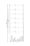
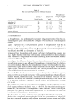
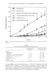
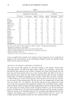
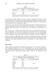
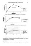
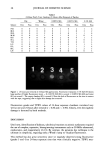
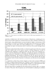
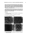
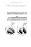
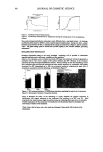
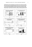
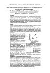
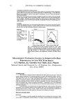
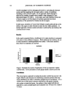
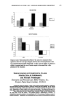
PYRANINE DETECTS INJURY TO SLS 35 fluorescence, with punctuate accentuation 4 = strong, bright fluorescence in large patches. Fluorescent photographs were also taken under two oppositely placed Wood's lamps (Spectroline ©, MB-100, 365 nm, Spectronics Corp., Westbury, NY) positioned 15 cm from the surface at a 45 ø angle. A Haze 2A Tiffen filter was placed in front of the camera lens. The film was 1600 ASA Kodak Ektachrome, processed at P2. TRANSEPIDERMAL WATER LOSS (TEWL) TEWL was measured 30 minutes after removal of the chambers, with the evaporimeter (EP2, Servo Med, Stockholm, Sweden) connected to a dedicated software. Measurements were conducted inside an environmental chamber at 21øC degrees and 41% relative humidity, after equilibration, following international guidelines for the use of this instrument (6). STATISTICS Significance was estimated by the Wilcoxon signed rank two-tailed test. RESULTS CORRELATION OF VISIBLE IRRITATION WITH FLUORESCENCE None of the one-hour patch tests, including 10% SLS, induced any visible reaction. However, with 10% SLS there was a non-significant increase in fluorescence when compared to water. With 24-hour exposures, only four of the 11 subjects showed a visible reaction to 0.025% SLS (grade 2) and 0.1% SLS (grades 2 and 3) one hour after removal. At 24 hours after removal of the chambers, two subjects with 0.025 % SLS and three with 0.1% SLS still had a mild erythema (Table I). The irritation was non-significant at any time compared with the water chamber. By contrast, strong fluorescence was observed (grades 3 and 4) in 100% of the sites exposed to 0.1% SLS (p 0.005, compared with all the other test sites), and in 63% of those exposed to 0.025% SLS (p 0.005, compared with water), one hour after removal. Fluorescence was still strong 24 hours after removing the chambers, although reaching significance only for 0.1% SLS (p = 0.001), in com- parison with water (Table I). No increase in fluorescence was observed with 0.005 % SLS (Figure 1). TEWL After the one-hour exposures, the TEWL values did not increase significantly. By contrast, with 24-hour patches TEWL values became significantly elevated (p 0.1), with all concentrations at one and 24 hours post removal (Figure 2). These increases were significant compared to water for 0.025% and 0.1% SLS (p 0.1). It should be noted that water also caused barrier damage compared to the empty chamber.

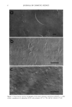
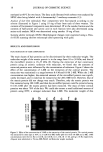
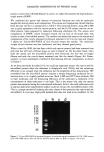
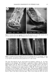
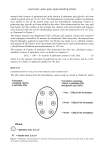
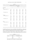
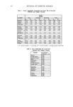
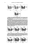
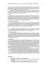
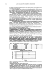
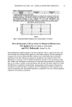
36 JOURNAL OF COSMETIC SCIENCE Table I 24-Hour Patch Tests: Grading 24 Hours After Removal of Patches Dry Water 0.005% SLS 0.025% SLS 0.1% SLS Subject Clin Fluo Clin Fluo Clin Fluo Clin Fluo Clin Fluo P.B. 0 0 0 2 0 2 0 2 1 4 M.S. 0 0 0 0 0 1 1 0 3 2 D.R. 0 0 0 2 0 2 0 3 1 4 M.H. 0 0 0 1 0 1 0 3 0 4 D.F. 0 0 0 0 0 2 0 4 1 3 N.L. 0 1 0 2 0 3 0 3 0 4 T.B. 0 2 0 0 0 0 0 0 0 3 L.C. 0 0 0 1 0 2 0 3 I 3 E.L. 0 0 0 0 0 1 3 1 3 4 B.C. 0 0 0 0 0 0 2 2 2 3 L.P. 0 0 0 0 0 0 0 0 0 3 12345 Figure 1. 24 hours post removal of 24-hour SLS applications. Fluorescent evaluation: 0.1% SLS (#3) shows large patches of bright fluorescence (score = 4). 0.025% SLS (#4) is scored 3.0.005% SLS (#2) and water (#1) are scored 1. The empty chamber (#5) is scored 0. Note the halo of fluorescence at the sites in contact with the tape, suggesting that a slight skin damage has occurred. Fluorescence grades and TEWL values of 24-hour exposure chambers correlated very well at one and 24 hours after removal (r = 0.86 and r = 0.94). Clearly, even the slightest damage is detected by both methods. DISCUSSION Until now, identification of dubious, subclinical reactions to anionic surfactants required the use of complex, expensive, bioengineering instruments such as 20 MHz ultrasound, conductance, and evaporimetry (2,3,5). By contrast, the pyranine dye technique is the ultimate in simplicity, requiring only a Wood's lamp to visualize fluorescence. This method has also great sensitivity since we regularly observed strong fluorescence (grades 3 and 4) at 24-hour exposure sites that were clinically negative. TEWL mea-
Purchased for the exclusive use of nofirst nolast (unknown)
From: SCC Media Library & Resource Center (library.scconline.org)