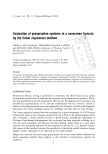
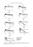
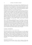
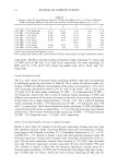
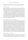
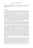
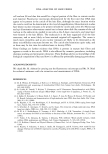
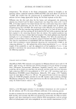
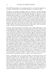
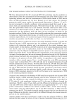
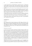
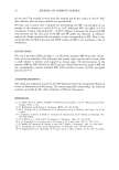
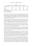
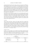
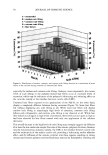
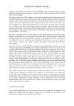
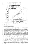
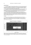
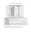
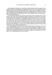
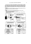
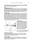
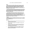
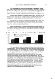
2002 ANNUAL SCIENTIFIC MEETING 109 Skin luminance and skin color were evaluated using a Chromameter. Redness, often a sign of inflammation was a measurement oferythema. There were no differences found between the three groups. There were unexpectedly high values in brightness with the young xerotics. The surface of their skin was uniformly covered with fine white scales. The elderly by contrast, was patchy and uneven yielding a low brightness value. Stratum corneum thickness was evaluated via shave biopsy. The horny layer was thickened in both xerotic groups. Average thickness in the elderly was 30+ layers, the young xerotics averaged 24 layers and the young normal have 18 or fewer layers. Skin hydration was measured via Corneometer, Novameter and via TEWL. The drier the skin the lower the Corneometer, and Novameter readings. The outer most corneocytes ofxerotic skin regardless of age are dried out, less hydrated and hold less water. They are brittle instead of soft and flexible. Dansyl chloride extinction of florescence results showed the elderly to be the first group to reach the point of patchy florescence, due to the large thick scales found on their skin. However the elderly were the last to reach final florescence extinction, due to the significantly slower rate of cell renewal. D a n sy 1 C h 1o rid e E x tin c tio n P a tte rn 30-• •ffilll P atc h y F Iou rescen ce l$ 1C om plete E xtin ction of Fluorescence 2 0 I 5 •.•:i ß ß ...., .. .:...:.........: .,:•. •... o ¾ o u n g, IN o rm a I ¾ o u n g, X erosis EIderly, X erosis This study helped to explain the biophysical differences ofxerotic skin with the young and elderly. Xerotic skin regardless of age showed little or no increase in redness when measured visually or with the chromamz.:er. This lack of redness indicates xerotic skin is not age dependant and is not an inflammatory condition. Laser Doppler studies showed a significant decline in the elderly. This decline is attributed to a reduction in blood vessels in the elderly. Fewer blood vessels result in a general lack of responsiveness to chemical irritants. Skin thickness increases with increasing age, clinically demonstrated by both a decrease in water loss as measured by TEWL and reduced reactivity to topical applied irritants. The elderly and the young xerotics showed and a decrease in skin hydration. Skin of both elderly and young xerotic groups was dried in appearanc, e and less hydrated•il'h a reduc, erl ahility to hold water• The harder function is compromised by the xerotic state regardless of age. Complete Dansyl Chloride extinction was slower for the young and old xerotics. This slower extinction rate indicates a thickening of the xerotic skin. The elderly rate however was much longer, attributable to the slower rate of cell renewal seen with age. Dry skin will continue as a vexing problem, but with continued studies such as this one, cosmetic chemists will be better able to provide long term effective relief.

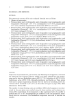
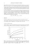
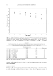
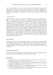
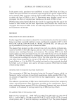
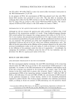
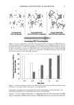
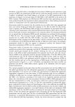
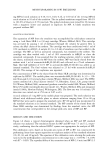
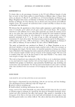
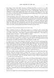
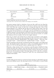
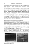
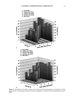
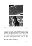
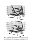
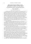
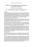
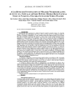
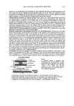
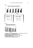
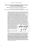
110 JOURNAL OF COSMETIC SCIENCE IDENTIFICATION OF ANOTHER BARRIER IN HUMAN STRATUM CORNEUM: THE WATER BARRIER Johann W. Wiechers', Ph.D., Joke A Bouwstra 2, Ph.D. and Anko de GraafF •Uniqema, PO Box 2, 2800AA Gouda, The Netherlands 2Leiden/Amsterdam Center of Drug Research, PO Box 9502, 2300 RA Leiden, The Netherlands Introduction: For more than two decades the intercellular lipid composition has been identified as the barrier to the influx of chemicals from the outside environment. But whilst this barrier does perform an excellent job in keeping the external world outside, we still lose considerable amounts of water every day via Transepidermal water loss. How can this barrier in one direction be so tough to cross but from the other direction not be watertight? Why do we not completely dehydrate in a dry environment? In order to address these rather fundamental questions, knowledge of the water distribution in human stratum comeurn (SC) at various relative humidities is required. In previous studies it has been hypothesized that water is partly absorbed in the corneocytes and remains partly in the intercellular regions in separate domains, i.e., phase separated from the lipid lamellar domains. Absorption of water into the comeocytes is expected to increase the volume of the comeocytes. But, since comeocytes are entirely surrounded by a crystalline monolaye r of lipids (lipids bound to the cornified envelope), a change in the lipid density of this monolayer is not expected to occur. Therefore, it is hypothesized that swelling of the comeocytes has to occur without inducing a strong change in the total surface area of the comeocytes. Methods: A new technique was developed that allowed the water distribution in the SC at various hydration levels to be studied: cryo-planing in combination with cryo scanning electron microscopy (Cryo- SEM) to obtain flat cryo-sections across the entire stratum comeum. This is an excellent method to determine both the distribution of water as a function of localization in the SC and the shape (that is the cross section) of the comeocytes as a function of the hydration level from one single image. SC was cut into sheets of 8 x 8 mm 2 and equilibrated over various salt solutions with known relative hurrfidities. After folding and embedding in tissue-freezing medium, the samples were rapidly frozen in liquid propane and sectioned in an ultramicrotome at a sample temperature of-90øC and freeze-dried for 3 minutes at the same temperature to obtain contrast and sputtered with a 5nm thick layer of platinum. Thereafter, they were subjected to SEM at a temperature of-190øC. Water that was present in the original SC sectioned samples will have sublimated during the freeze-drying procedure from the flat surface, creating a relief (contrast) that can be identified as a lighter surface in the cryo-SEM's. The technique is illustrated in Figure I. Numerical parameters such as average cell thickness, circumference as well as the enclosed surface area could be calculated from the digital cryo- SEM's. Figure 1 Schetnatic presentation of the sequential events of the Cryo-Scan ing electron microscolby technique Cryo-planing in combination with cryo-SEM V._•.•Freshlobtainedsurfaces tissue fr.
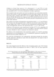
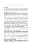
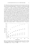
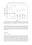
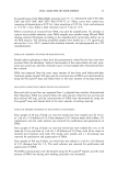
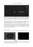
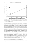
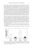
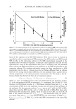
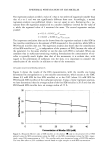
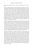
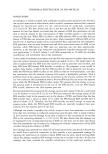
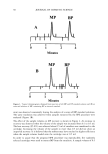
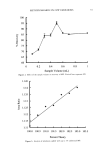
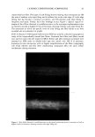
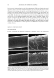
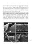
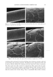
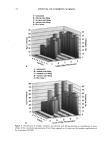
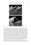
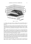
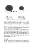
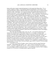
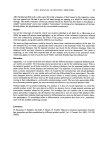
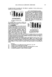
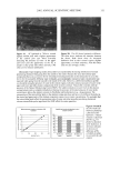
Embedding eezing C•yo-freezing planing Dehydration Cryo-SEM Results: Using this new method, it was discovered that water in the SC is neither homogeneously distributed, nor gradually increasing with depth. Dry skin and skin hydrated to 17% w/w was characterized by low contrast and cells show many undulations, particularly close to the comeocyte cell ends. At 57- 87%w/w water content the hydration level in the central part of SC is higher than in the superficial and deeper cell layers. Water domains are mainly present within the comeocytes and not in the intercellular regions (see Figure 2). At a very high hydration level (300 %w/w), the comeocytes are strongly swollen except for the deepest cell layers adjacent to the viable epidermis. The comeocytes in these layers are not 17%w/w and 300%w/w the average cell thickness increases linearly with the hydration level suggesting that swelling of cells mainly occurs in the direction perpendicular to the skin surface. At an increased hydration level, the comeocyte envelope surrounds the cell content more efficiently (see Figure 3) compensating for the increased cell volume. The changes in stratum corneum morphology with increasing water level have also been observed in dermatomed skin indicating that the observations are not simply an artifact due to the lack of underlying tissue.
Purchased for the exclusive use of nofirst nolast (unknown)
From: SCC Media Library & Resource Center (library.scconline.org)