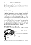
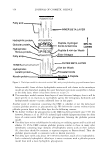
328 JOURNAL OF COSMETIC SCIENCE to accommodate the close packing of the three chains. The extended conformation of the individual polyproline II-like chains in the triple helix is stabilized by the high level of occurrence of the amino acids praline and 4-hydroxyproline (Hyp). Hyp, which is post-translationally modified from praline, confers a greater stability than praline in the Y position (6) and is a characteristic amino acid marker of collagen. In tissue, the collagen molecules are crosslinked to form an extended network that provides the tissue with strength and durability. These crosslinks are specific and use short, non-triple helical peptides, the telopeptides, at the ends of the triple-helical domain of each collagen molecule (7). Bovine collagen has been a common source for cosmetic applications. However, more recently, collagens from other species of origin have also become commercially available. Collagens from alternative species may provide a benefit in that potential zoonoses, particularly transmissible spongiform encephalopathies, are less likely from sources such as chicken and fish. However, collagens from various species may differ in their prop erties. For example, they may have different thermal stabilities (8), which could affect formulation or the shelf life of products. In the present study we have examined the suitability of a range of tests, using a selection of commercially available collagen samples. These samples illustrate a selection from the range of materials available and allow commentary on the information that each test provides. Some tests, those found in water analysis standards (9), for example, are readily available from many analytical laboratories, whereas other tests, which may be critical for providing discrimination between samples, may require the involvement of more spe cialized laboratories. MATERIALS AND METHODS COLLAGEN SAMPLES Commercial collagen samples suitable for cosmetic application were obtained from the following sources: Collasol® was from Croda Chemicals Ltd (Humberside, UK). CLR Collagen® was from Chemisches Laboratorium Dr. Kurt Richter Gmbh (Berlin, Ger many). AteloHelogen® was a gift from the manufacturer (Meddicoll, North Ryde, Australia). Soluble collagen samples were also prepared in our laboratory from fresh, minced dermis (bovine, porcine, and chicken, obtained from local abattoirs) by pepsin digestion using 1 mg/ml pepsin in 100 mM acetic acid adjusted to pH 2.5 with HCl (10). Soluble collagen from diced fish skin (Blue Grenadier (Macruronus sp.) and Nile perch (Tilapia sp.) obtained from a local supplier) was extracted using 0.1 mg/ml pepsin in 100 mM acetic acid adjusted to pH 2.5 with HCl. All collagens were purified by differential NaCl fractionation at pH 2.5 and then at pH 7.4 (11). Separation of type I and III collagens was by rapid ammonium sulfate fractionation (11). METHODS Solution analysis. Analyses for ash, arsenic, and heavy metals (as lead) were performed by a certified external analytical laboratory (MGT Environmental Consulting Pty. Ltd., Oakleigh, Australia) following standard procedures (9). pH was determined using a

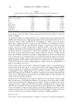
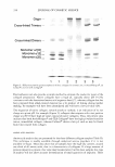
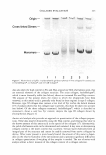
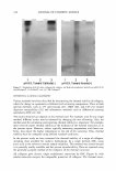
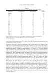
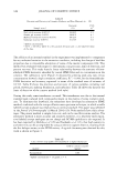
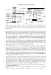
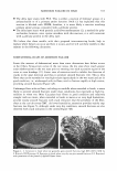
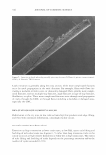
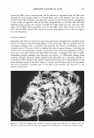
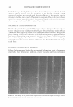
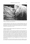
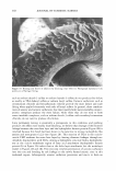
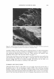
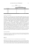
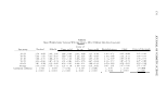
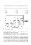
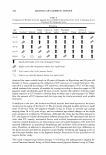
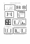
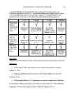
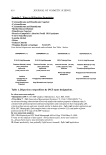
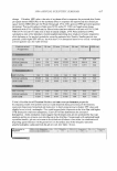
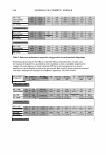
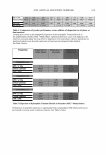
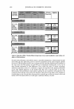
COLLAGEN EVALUATION 329 Radiometer PHM240 instrument (Copenhagen, Denmark). Conductivity was deter mined using a Radiometer CDM3 instrument. Water regain. Collagen samples were dried by lyophilization in preweighed bottles with stoppers and then held over silica gel for more then 72 h. Samples were then stoppered and reweighed. They were then transferred and opened in an environment of 32% constant relative humidity, maintained over saturated CaC12 solution. After equilibra tion for more than 48 h, the samples were stoppered and reweighed. Water regain by the dried samples was determined, and expressed as a percentage of the dry weight. Spectroscopy. For UV/visible spectroscopy, spectra were collected from 400 nm to 650 nm on a Shimadzu UV-265 recording spectrometer using cells of 10-mm path length on collagen solutions as supplied or after dilution to equal concentrations of 1 mg/ml with milliQ water. For IR spectroscopy, collagen samples were lyophilized and then examined using a Perkin Elmer 2000 Autolmage spectrometer with the ATR accessary. Alterna tively, samples may be analyzed as KBr disks. Electrophoresis. Collagen samples were analyzed by sodium dodecyl sulfate-polyacrylamide gel electrophoresis (SDS-PAGE) (12), using 5% (w/v) running gels. Prior to analysis, collagen samples in sample buffer were neutralized, if necessary, with 2 M Tris and then heated at 100°C for 2 min. Separation of the al(I) and al(III) chains was by reduction with 2-mercaptoethanol during interrupted electrophoresis (13 ). Collagen samples were also examined by a non-denaturing, lactic acid buffer system, pH 3 .1 (14), using 4% running gels. Estimation of the isoelectric point, pl, was by examination of the direction of electrophoretic mobility in gel electrophoresis in 5% running gels in a Tris/Borate pH 8.9 system (15). Amino acid analysis. Lyophilized collagen samples were hydrolyzed in vacuo by constant boiling (5.8 M) of HCl containing 0.1 % phenol in a Waters PicoTag system at 108°C for 20 h. After drying in vacuoJ hydrolysates were examined on a Waters HPLC system with ninhydrin detection. Differential scanning calorimetry. Collagen melting temperatures (T m ) were determined by DSC using a Mettler Toledo DSC 3000 instrument (Mettler, Schwerzenbach, Switzer land). Collagen samples were prepared at about 5 mg/ml, and equilibrated in 50 mM acetic acid, pH 2.8, by dialysis. Samples, about 100 mg of solution, were examined at a temperature increase of 1 ° C/min. Collagen T m values are given as the temperature at the mid-point of the thermal transition. Scanning electron microscopy. Collagen samples as provided, or after dilution to equal concentrations of 3 mg/ml with milliQ water, were coated on clean glass coverslips and air dried at room temperature in the clean air flow of a laminar flow hood. Dried samples were then gold coated and examined in a Philips XL-30 microscope. RESULTS AND DISCUSSION GENERAL SOLUTION PROPER TIES There are a wide variety of solution measurements that can be readily made on collagen preparations (Table I). These include analysis for toxic metal components such as arsenic and lead, whose presence would make a preparation unsuitable for cosmetic application.
Purchased for the exclusive use of nofirst nolast (unknown)
From: SCC Media Library & Resource Center (library.scconline.org)