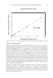
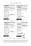
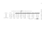
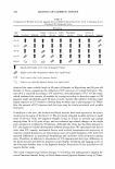
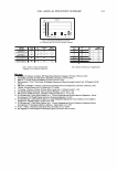
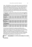
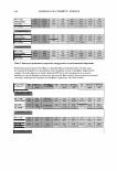
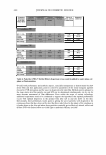
ASSESSMENT OF OCULAR IRRITANCY 319 irritation was determined by ascertaining from the subject any experiences of ophthalmic irritation (i.e., stinging, burning, itching, dryness, and/or foreign body sensation) at the time of the specified examination. Subjects were examined for evidence of excessive lacrimation. Each subject's upper and lower eyelids, specifically the lid margins, were examined for evidence of redness, scaling, swelling, and/or excessive secretions of the meibomian gland orifices. The palpebral and bulbar conjunctivae were examined and scored for redness, inflammation, and follicular and/or papillary reactions. The cornea was examined for evidence of any neovascularization, edema, infiltrates, opacities, and/or epithelial defects. To assess fluorescein staining patterns, a Fluorets sterile ophthalmic strip (fluorescein sodium BP, Smith & Nephew) was inserted into the inferior cul de sac of each eye after a small amount of Dacriose sterile irrigating solution had been dropped onto the Fluorets® strip. The integrity of the palpebral and bulbar conjunctivae, corneal epithelium, and caruncle were then evaluated with a Haag-Streit Bern model Z 2982A slit lamp biomicroscope, and the tear film break-up time was assessed. DATA ANALYSES The grading scale, designed by Bruce E. Kanengiser (3), for all parameters was 0--4 (0 = normal, 1 = trace, 2 = mild, 3 = moderate, and 4 = severe), with the exception of fluorescein ophthalmic staining, in which a 14-point scale was employed to evaluate the integrity of the palpebral and bulbar conjunctivae, corneal epithelium, and caruncle (Table II). Additionally, tear film break-up time was assessed by determining the time that elapsed before the first hole (dry spot) appeared in the corneal fluorescein layer(::::::10 seconds is normal). Microsoft Excel (2000) and SigmaStat/SigmaPlot Version 5 were used to compile and statistically analyze the data. Student's t-test and ANOV A and correlation (r-value) tests were performed. Statistical significance was declared for all Table II Palpebral and Bulbar Conjunctival, Caruncular, and Corneal Fluorescein Ophthalmic 14-Point Area Staining Scale (3) Ocular irritancy No ocular irrirancy Mild ocular irritancy range Moderate ocular irri tancy range Severe ocular irritancy range Level 0 2 3 4 5 6 7 8 9 10 11 12 13 Description (% staining in quadrant) No staining 0 and ::=:::10% 10% and ::=:::20% 20% and :2::30% 30 and ::=:::40% 40% and ::=:::so% 50% and :2::60% 60% and ::=:::70% 70% and ::=:::so% 80% and :2::90% 90% and :2::100% Mild superficial tissue abrasion Moderate superficial tissue abrasion Severe superficial tissue abrasion Density grading scale: 1 = occasional, scattered punctate staining 2 = more uniform pattern of diffusely scattered punctate staining 3 = dense foci of punctate staining within the areas of diffuse punctate staining 4 = general pattern of dense punctate staining.

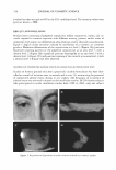
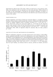
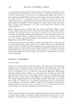
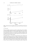
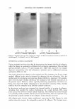
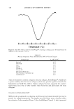
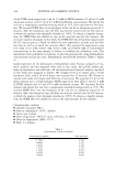
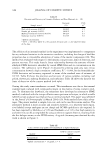
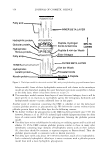
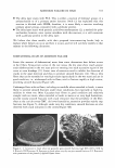
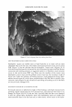
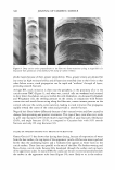
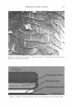
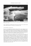
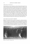
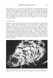
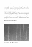
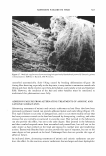
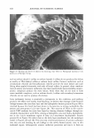
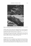
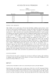
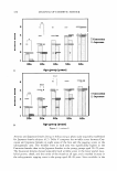
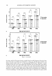
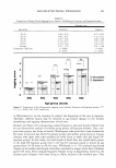
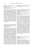
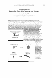
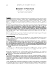
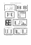
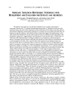
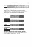
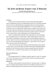
320 JOURNAL OF COSMETIC SCIENCE p-values less than or equal to 0.05 at the 95% confidence level. The intensity values were given as means ± SEM. RESULTS AND DISCUSSION Ocular tissues consisting of palpebral conjunctiva, bulbar conjunctiva, cornea, and ca runcle respond to cosmetic exposure with different severity, pattern, and/or onset of symptoms, with respect to inflammation, abnormalities, and/or observed tissue abrasion. Figure 1 depicts ocular irritation induced by instillation of a cosmetic or consumer product. Moderate inflammation of the conjunctivae at a level 3 (Figure lA) punctate fluorescein staining patterns of the palpebral conjunctivae at an area level 3 with a density level 2 (Figure lB) superficial punctate keraropathy at an area level 3 with a density level 3 (Figure lC) and punctate staining of the caruncle at an area level 4 with a density level 3 (Figure 1D) were observed. INCIDENCE OF UNEXPECTED ADVERSE EVENTS IN HUMAN OCULAR INSTILLATION TESTS Studies on humans proceed only after a potentially suitable formulation has been iden tified as a result of irritation tests in animals and in vitro (4), minimizing the potential of unexpected adverse events during in vivo studies. The frequency of occurrence of adverse events was minimal in human ocular instillation studies. Of 205 human subjects who participated in ocular instillation studies from 1998 to 2003, only one subject Figure 1. Representative illustrations of ocular irritation induced by a cosmetic product.
Purchased for the exclusive use of nofirst nolast (unknown)
From: SCC Media Library & Resource Center (library.scconline.org)