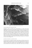
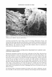
]. Cosmet. Sci., 55, 351-371 Quly/August 2004) Failure of intercellular adhesion in hair fibers with regard to hair condition and strain conditions CLARENCE ROBBINS, HANS-DIETRICH WEIGMANN, SIGRID RUETSCH, and YASH KAMATH, 12425 Lake Ridge Circle, Clermont, FL 34711 (C. R.), and Textile Research Institute, PO Box 625, Princeton, NJ 08540 (H.-D. W., S. R., Y. K.). Accepted for publication March 31, 2004. Synopsis Although adhesion failure in hair fibers can occur inside cells, it occurs more frequently in the cell membrane complex (CMC), often involving the rupture of interlayer bonds. Therefore, a model of the CMC is presented, based on prior research in which we propose interconnecting bonds between the layers to assist in our interpretation of hair-fracturing mechanisms for cuticle chipping, deep transverse cuticle cracks, cracks during heat drying, scale lifting by surfactants, and catastrophic failure. Failure in the wet state generally involves hydrophilic layers, e.g., the contact zone of the CMC or the endocuticle or bonding to these hydrophilic layers, whereas failure in the dry state generally involves bonding between hydrophobic layers, e.g., beta-delta failure. Chemical damage by perms, bleaches, and sunlight, by breaking specific chemical bonds, influences the sites of initial failure and increases the number of routes for crack propa gation, leading to more complex fracture patterns. INTRODUCTION Failure of adhesion can occur at different sites in hair fibers, depending on the condition of the hair and the conditions of strain that produce failure. The condition of the hair is determined primarily by prior treatments and fiber twists and/or flaws, including whether or not it contains a medulla and whether it has been chemically treated or exposed to sunlight, and the kind and amount of sorbed materials. The conditions of strain include stretching, bending, twisting, percentage deformation, the employment of heat such as heat drying, and water content or percent relative humidity. The cosmetic importance of adhesion failure resides in the fact that it controls hair breakage and fragmentation, actions that are so important to the look, the feel, and the condition of the hair. Fragmentation of hair is determined primarily by chemical treat ments to the fibers, including sunlight exposure, the conditions of deformation, and abrasive actions that are employed in everyday grooming actions (1). It is the combi nation of these actions at high water content that results in the most severe hair erosion, especially when combined with repetitive actions, as in extension cycling, fatiguing, and abrasion to break. 351

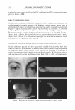
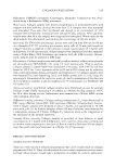
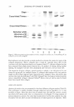
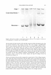
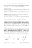
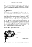
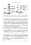
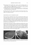
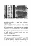
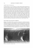
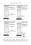
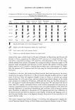
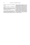
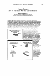
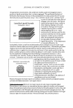
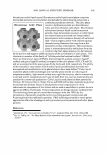
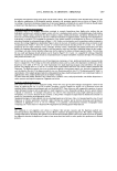
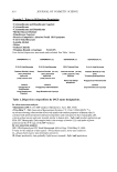
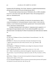
352 JOURNAL OF COSMETIC SCIENCE Although failure can occur inside cuticle and cortical cells, the cell membrane complex (CMC) is involved in several different types of adhesion failure and frequently is the origin of fracture and crack propagation. Therefore, a model of the CMC is provided in this paper to help explain where and how failure occurs under several different condi tions. HAIR FIBER STRUCTURE The CMC is an interconnecting unit consisting of three different types that are very similar structurally (see Figure 1). The CMC between cuticle cells is called cuticle cuticle CMC, at the junction of the cuticle and the cortex is the cuticle-cortex CMC, and the CMC between cortical cells is called cortex-cortex CMC. Of these three structures the cuticle-cuticle CMC and the cortex-cortex CMC have been studied to the greatest extent. The subtle known differences in these structures are that the cuticle-cuticle CMC contains 18-methyl eicosanoic acid (18-MEA) in one of its beta layers (outer beta layer, the one immediately on top of cuticle cells), while the other two CMCs do not contain this specific fatty acid, but consist of straight-chained fatty acids from Cl2 through Cl8 and oleic acid in place of this unique branched fatty acid (see Figure 2A). 18-MEA is distinctive in its role in the cuticle-cuticle CMC (2), which will be explained later in this paper. Since 18-MEA is a C2 l fatty acid and exists only in the outer beta layer of the cuticle-cuticle CMC (3) and is replaced by shorter-chain fatty acids in the cortex-cortex CMC, the cuticle-cuticle CMC should be thicker than the cortex-cortex CMC. The cuticle-cuticle CMC has been shown to link fatty acid (18-MEA) to cuticle cell mem branes in the outer beta layer (Figure 2A) through thioester linkages (4), while it would appear from the work of Bryson et al. (5) that fatty acids in the cortex-cortex CMC are linked to the hydrophobic keratin protein layer through ester linkages. THE STRUCTURE OF THE CUTICLE-CUTICLE CMC Nearly three decades ago, Fraser and coworkers (6) described the CMC as consisting of 9!1i1 1ll!lltll lill!II 11 '81!111! 11111 •111• •■111811 8 ..........Cortex-Cortex CMC . . ....................·· ········Cuticle-Cortex CMC . .... .......... ....... .. · ········cuticle-Cuticle CMC Figure 1. The CMC, a structure where failure tends to occur. There are three types of CMC in human hair that are structurally different.
Purchased for the exclusive use of nofirst nolast (unknown)
From: SCC Media Library & Resource Center (library.scconline.org)