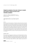
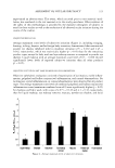
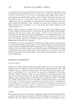
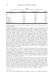
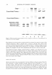
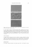
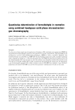
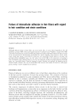
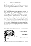
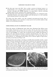
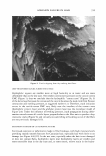
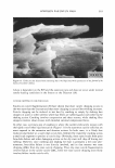
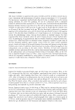
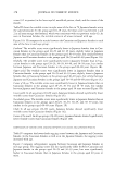
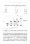
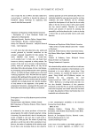
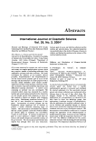
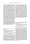
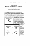
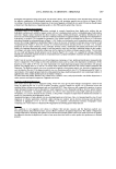
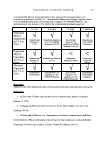
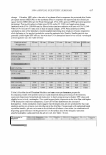
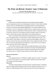
352 JOURNAL OF COSMETIC SCIENCE Although failure can occur inside cuticle and cortical cells, the cell membrane complex (CMC) is involved in several different types of adhesion failure and frequently is the origin of fracture and crack propagation. Therefore, a model of the CMC is provided in this paper to help explain where and how failure occurs under several different condi tions. HAIR FIBER STRUCTURE The CMC is an interconnecting unit consisting of three different types that are very similar structurally (see Figure 1). The CMC between cuticle cells is called cuticle cuticle CMC, at the junction of the cuticle and the cortex is the cuticle-cortex CMC, and the CMC between cortical cells is called cortex-cortex CMC. Of these three structures the cuticle-cuticle CMC and the cortex-cortex CMC have been studied to the greatest extent. The subtle known differences in these structures are that the cuticle-cuticle CMC contains 18-methyl eicosanoic acid (18-MEA) in one of its beta layers (outer beta layer, the one immediately on top of cuticle cells), while the other two CMCs do not contain this specific fatty acid, but consist of straight-chained fatty acids from Cl2 through Cl8 and oleic acid in place of this unique branched fatty acid (see Figure 2A). 18-MEA is distinctive in its role in the cuticle-cuticle CMC (2), which will be explained later in this paper. Since 18-MEA is a C2 l fatty acid and exists only in the outer beta layer of the cuticle-cuticle CMC (3) and is replaced by shorter-chain fatty acids in the cortex-cortex CMC, the cuticle-cuticle CMC should be thicker than the cortex-cortex CMC. The cuticle-cuticle CMC has been shown to link fatty acid (18-MEA) to cuticle cell mem branes in the outer beta layer (Figure 2A) through thioester linkages (4), while it would appear from the work of Bryson et al. (5) that fatty acids in the cortex-cortex CMC are linked to the hydrophobic keratin protein layer through ester linkages. THE STRUCTURE OF THE CUTICLE-CUTICLE CMC Nearly three decades ago, Fraser and coworkers (6) described the CMC as consisting of 9!1i1 1ll!lltll lill!II 11 '81!111! 11111 •111• •■111811 8 ..........Cortex-Cortex CMC . . ....................·· ········Cuticle-Cortex CMC . .... .......... ....... .. · ········cuticle-Cuticle CMC Figure 1. The CMC, a structure where failure tends to occur. There are three types of CMC in human hair that are structurally different.

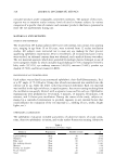
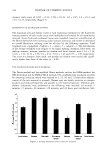
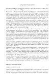
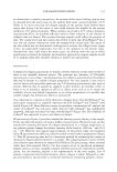
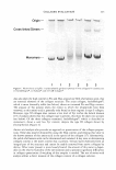
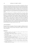
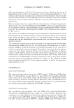
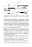
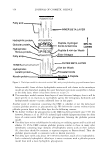
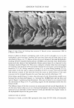
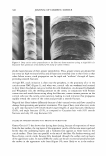
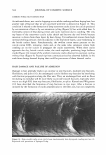
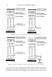
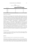
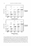
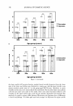
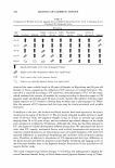
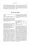
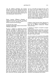
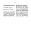
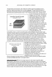
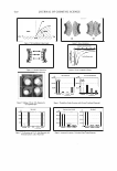
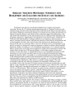
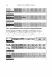
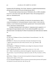
ADHESION FAILURE IN HAIR 353 = f Fiber Surface -------: r ! r !- � INNER BETA LAYER 008fOO_p_ep-ti-de-Hy_d_rog_ e_n---,-r- Hydrophilic Protein(�/ bonds&. Salt links DEL TA "Contact Zone" � Globular Protein (e)�OooJBoi LAY l ER Hydrophobic -Peptide & van der W aals fibrous protein (d) � - lB-MEA ( c� f f ---'------Van der W a :: ER B ET A L AYER Hydrophobic . __ _ I keratin protein �.l___J)isulfide �ioesterbonds & Peptide Keratin protein (a}-- bonds A LAYERS of CMC INTERLAYER BONDING Hydrophil�j_:i, �tide� H dr�g n- � protein f O O nD o Bonds & Salt Links : Globular Protein e-0ao O�eptide & : Hydrophobic � van der Waals 1 fibrous protein d f-� �� � -van der Waals : 18-MEA c-f f f"•u•L:: T _ -/-��A _ Th1oester ,, Hydrophobic � ,, ,, Keratin Protein b- nal.clx.il2nac: Disulfkfe � & Peptide Keratin Protein a- ' A-Layer--- ◄- - - - - - - -'PEROXIDE B Figure 2. A. The monolayer model for the cuticle-cuticle CMC, with principle bonding proposed between layers. The inner beta layer is the "lower" and the outer is the "upper" beta layer. B. The monolayer model for the cuticle-cuticle CMC (lower half) and its principle interlayer bonds, with some primary sites for chemical attack. two lipid layers (called inert beta layers) separated by intercellular cement, the delta layer (Figure 2A). Several important developments have occurred since Fraser's description of the CMC that have further improved our understanding of this important structure. Among these important developments are the work of Rivett and coworkers (3 ,4) and Bryson and coworkers (5 ). Two different models are presented in this paper in which this prior research is taken into consideration to provide a more detailed chemical represen tation of the CMC. In our models, we are proposing the interconnecting bonds between the different layers of the CMC to assist in our interpretation of hair-fracturing mecha nisms. Both of our models consist of protein and fatty acid layers. Model 1 (Figure 2A,B) contains monolayers in the beta layers, while model 2 (Figure 3) contains bilayers in the fatty acid domains consistent with Fraser's original concept. We are also suggesting a change in terminology. Up to this point, the beta layer immediately on top of each cuticle cell has been called the upper beta layer and the one beneath each cuticle cell has been called the lower beta layer. This terminology is confusing, especially for schematics of the CMC as in Figures 2A,B and 3. Therefore, we propose that the beta layer that sits on top of each cuticle cell and often becomes the outer hair surface be called the outer beta layer and the one immediately beneath each cuticle cell be called the inner beta layer (see Figures 2A,B and 3), and this terminology will used throughout this manuscript. Figure 2B shows only one-half of the CMC structure for the monolayer model and depicts some of the primary sites for chemical attack by permanent waves, bleaches, and sunlight. The top half of the CMC is not depicted in this figure because it is similar to the lower half, with the exception that it does not contain 18-MEA but contains saturated fatty acids (Cl2-Cl8) and oleic acid in place of 18-MEA. Our preference is for the monolayer model over the bilayer model for the following reasons: • Swift (2) has pointed out that a monolayer model fits better from the point of view of CMC measurements. • If the beta layers are monolayers, then 18-MEA is linked to the delta layer through short hydrophobic side chains rather than through ester or thioester bonds (in the
Purchased for the exclusive use of nofirst nolast (unknown)
From: SCC Media Library & Resource Center (library.scconline.org)