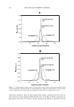
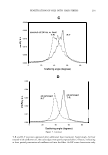
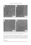
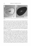
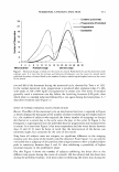
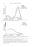
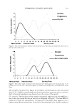
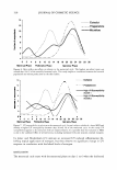
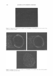
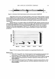
320 JOURNAL OF COSMETIC SCIENCE estrogens have been reported to improve the extracellular matrix responsible for the tone and appearance of the skin (26). The lowest production of progresterone and estrogen was on the first few days of the cycle, when skin appeared to be the most dry. In addition, skin surface lipids appeared to be highest on days 16-20 of the hormonal cycle. The pattern of skin oiliness appears to follow the estrogen release pattern, indicating a possible effect of this hormone on skin surface lipids. The highest microbial count was around days 16 to 22 of the monthly hormonal cycle. The microflora resident on human skin show great inter-individual and intra-individual differences. It is essentially composed of micrococci, staphylococci, and aerobic and anaerobic coryneforms, as well as pityrosporum species, which, in accordance with the different environment in the different regions of the body, are in a steady state. With increasing age, human skin microflora undergo qualitative changes: the streptococci, which are found in infants, disappear and coryneform bacteria occur, which are mainly responsible for odor production (27). Human "native" intracellular sebum, before se cretion, is composed of squalene, waxes, and triglycerides. Once secreted, the sebum is colonized by various xenobiots whose development is controlled by several defensive humoral mechanisms and by contact with ambient oxygen. Oxygen and microorganisms transform "native" sebum, the lysis of triglycerides to fatty acids (28). A correlation between bacterial population and sebum production has been implied (29) and is clearly visible in this study. Several subjects exhibited a lower MED and thereby a higher UV susceptibility between days 20 and 28 of the menstrual cycle. Studies conducted by Jemec and Heidenheim (23) indicate an increased UV-induced inflammation following topical application of estro gen, but they observe no significant change in UV response in correlation with the blood levels of estrogen. In this study, a weak barrier follows the maximum systemic production of estradiol and progesterone. In this study, UV susceptibility appears to be concurrent with barrier condition. Al though epidermal stratification and the formation of a stratum corneum has been hy pothesized to provide protection against UV-B radiation (30-31) the differences in UV-B transmission in both stratum corneum and epidermis are too small to account com pletely for variation in MED at the different times of the cycle. It is possible that this variation in MED is due to the combined effect of several factors, including hormonal levels and stratum corneum integrity. REFERENCES (1) E. Sbano, V. Altamura, F. Galasso, and C. Capilungo, Sexual hormones and acne in adolescent women, G. Ital. Dermatol. Venereal., 125, 363-367 (1990). (2) D. M. Lawrence, M. Katz, T. W. Robinson, M. C. Newman, H. H. McGarrigle, M. Shaw, and G. C. Lacheli'n, Reduced sex hormone binding globulin and derived free testosterone levels in women with severe acne, Clin. Endocrinol., 15, 87-91 (1981). (3) R. Linse and J. Hadlich, Dermatitis as an expression of progesterone hypersensitivity, Zentralbl. Gynakol., 100, 926-930 (1978). (4) C. Eisenbeiss, J. Welzel, and W. Schmeller, The influence of female sex hormones on skin thickness: Evaluation using 20 MHz sonography, Br.]. Dermatol., 139, 462-467 (1998). (5) J. Harvell, I. Hussona-Saeed, and H. I. Maibach, Changes in transepidermal water loss and cutaneous blood flow during the menstrual cycle, Contact Dermatitis, 27, 294-301 (1992). (6) L. Bungum, K. Kvernebo, P. Oian, and J.M. Maltau, Laser doppler-recorded reactive hyperaemia in the forearm skin during the menstrual cycle, Br.]. Obstet. Gynaecol., 103, 70-75 (1996).

HORMONAL CHANGES AND SKIN 321 (7) M.A. Giamberardino, K. J. Berkley, S. Iezzi, P. De Bigontina, and L. Vecchiet, Pain threshold variations in somatic wall tissues as a function of menstrual cycle, segmental site and tissue depth in non-dysmenorrheic women, dysmenorrheic women and men, Pain, 71, 187-197 (1997). (8) T. Agner, P. Damm, and S. 0. Skouby, Menstrual cycle and skin reactivity,]. Am. Acad. Dermatol., 24, 566-570 (1991). (9) S. C. Chattoraj and N. B. Watts, Endocrinology In Fundamentals of Clinical Chemistry, N. W. Tietz, Ed. (W. B. Saunders, Philadelphia, 1987), pp. 575. (10) J. Pinnagoda, R. A. Tupker, T. Agner, and]. Serup, Guidelines for trans epidermal water loss (TEWL) measurement, Contact Dermatitis, 22, 164-178 (1990). (11) G. Grove, Physiologic changes in older skin, Clin. Geriatr. Med., 5, 115-125 (1989). (12) P. M. Elias, Epidermal lipids, barrier functions and desquamation, J. Invest. Dermatol., 80, 44s--49s (1993). (13) P. Frosch and A. M. Kligman, "Recognition of Chemically Vulnerable and Delicate Skin," in Principles of Cosmetics for the Dermatologist, P. Frost and S. N. Horwitz, Eds. (C. V. Mosby, St. Louis MO, 1982), pp. 287-296. (14) H. Tagami, "Measurement of Electrical Conductance and Impedance," in Handbook of Non-Invasive Methods and the Skin, J. Serup and G. B. E. Jemec, Eds. (CRC Press, Boca Raton, Florida, 1995), pp. 159-164. (15) A. 0. Barel and P. Clarys, "Measurement of Electrical Capacitance," in Handbook of Non-Invasive Methods and the Skin, J. Serup and G. B. E. Jemec, Eds. (CRC Press, Boca Raton, Florida, 1995), pp. 165-172. (16) W. J. Cunliffe, J. N. Kearney, and N. B. Simpson, A modified photometeric technique for measuring sebum excretion rate,]. Invest. Dermatol, 75, 396 (1980). (17) R. R. Marples, "Newer Methods of Quantifying Skin Bacteria," in Skin Microbiology Relevance to Clinical Infection, H. Maibach and R. Aly, Eds. (Springer-Verlag, New York, 1981), pp. 45--49. (18) K. T. Holland, "Microbiology of Acne", in Acne, W. J. Cunliffe, Ed. (Martin Dunitz Ltd, London, 1989), pp. 178. (19) M. J. Pelczar, E. C. S. Chan, and N. Krieg, "The Microscopic Examination of Microorganisms," in Microbiology, Part 1: Introduction to Microbiology, 5th ed., M. J. Pelczar, E. C. S. Chan, and N. Krieg, Eds. (McGraw-Hill, New York, 1986), pp. 66-67. (20) Sunscreen Drug Products for Over the Counter Human Use, Department of Health Education and Welfare, Food and Drug Administration, Federal Register, 64, no. 98, 27666-27693 (May 21, 1999). (21) K. Hoffmann, K. Kaspar, G. von Kobyletzki, M. Stucker, and P. Altmeyer, UV transmission and UV protection factor (UPF) measured on split skin following exposure to UVB radiation-Correlation with the minimal erythema dose (MED), Photodermatol. Photoimmunol. Photomed., 15, 133-139 (1999). (22) P. J. Toot, E. S. Surrey, and J. K. H. Lu, "The Menstrual Cycle, Ovulation, Fertilization, Implantation and the Placenta," in Essentials of Obstretics and Gynecology, Second Edition, E. N. Hacker and J. G. Moore, Eds. (W.B. Saunders, Philadelphia, 1992) pp. 36-45. (23) G. B. Jemec and M. Heidenheim, The influence of sex hormones on UVB induced erythema in man. ]. Dermatol, Sci., 9, 221-224 (1995). (24) D. Kalogeromitros, A. Katsarou, M. Armenaka, D. Rigopoulos, M. Zapanti, and I. Stratigos, Influence of the menstrual cycle on skin-prick test reactions to histamine, morphine and allergen, Clin. Exp. Allergy., 25, 461--466 (1995). (25) M. J. Thornton, The biological actions of estrogens on skin, Exp. Dermatol., 11, 487-502 (2002). (26) M. P. Brincat, Hormone replacement therapy and the skin, Maturitas, 29, 107-117 (2002). (27) H. C. Korting, A. Lukacs, and 0. Braun-Falco, Microbial flora and odor of the healthy human skin Hautarzt, 39, 564-568 (1988). (28) D. Saint-Leger. Normal and pathologic sebaceous function: Research in a shallow milieu? Pathol. Biol., 51, 275-278 (2003). (29) J. N. Kearney, E. Ingham, W. J. Cunliffe, and K. T. Holland, Correlations between human skin bacteria and skin lipids, Br.]. Dermatol., 110, 593-599 (1984). (30) E. Corsini, N. Sangha, and S. R. Feldman, Epidermal stratification reduces the effects of UVB (but not UVA) on keratinocyte cytokine production and cytotoxicity, Photodermatol. Photoimmunol. Photomed., 13, 147-152 (1997). (31) W. A.G. Bruls, H. Weelden, and]. C. Van Der Leun, Transmission of UV-radiation through human epidermal layers as a factor influencing the minimal erythemal dose, Photochem. Photobiol. 1 39, 63-67 (1984).
Purchased for the exclusive use of nofirst nolast (unknown)
From: SCC Media Library & Resource Center (library.scconline.org)