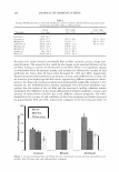
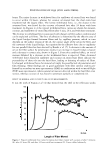
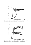
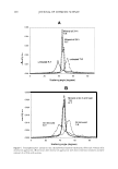
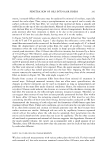
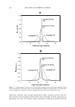
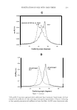
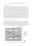
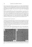
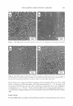
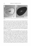
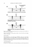
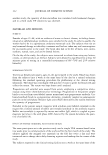
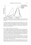
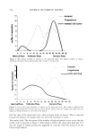
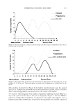
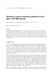
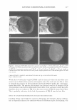
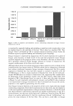
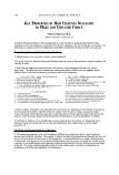
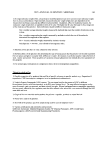
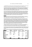
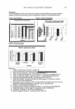
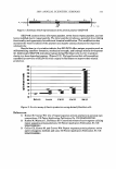
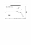
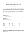
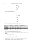
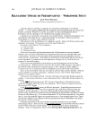
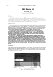
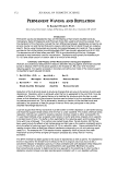
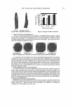
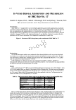
348 JOURNAL OF COSMETIC SCIENCE PROTECTING THE SKIN AGAINST OZONE James V. Gruber1, Ph.D., Abdullatif Tay1, Ph.D. and Robert Holtz2 1 Arch Personal Care Products, L.P., 70 Tyler Place, South Plainfield, NJ 07080 2 Bioinnovation Laboratories, Inc., 321 N Central Expressway, Suite 256, McKinney, TX Introduction Ozone (03) can be considered to be a transitory mixture of oxygen (02) and singlet OX)'gen c1 O:) and is an e,-1remely powerful, non-radical oxidizing agent.1 It has been well documented that ozone will readily kill microorganisms and it is frequently used as a sterilizing agent for this purpose. Unfortunately, ozone is also the most significant contaminate in urban air pollution and when local governments speak of pollution alerts, they arc typically talking about unhealthy levels of ozone. 2 Typical levels of ozone that a1 recorded in urban enYironments can range between 0.2 and 1.2 ppm with the higher leYels being considcre a respiratory threat to people with impaired breathing. 2 Ozone ravages the human respiratory system and also is now identified as a important source of oxidath·e stress and reactive oxygen species in the skin_ 3 · 5 In particular, ozone has been identified as a source of depletion of cutaneous vitamins3 and lipids4·5. In addition, it is well established that ozone can damage proteins and nucleic acids although such studies on skin have been sparse. 6 More recently. it was suggested that atmospheric ozone may adversely effect the immune response in frogs, in particular the ability of pulmonary macrophages to function correctly, and that this effect may partly account for the decline in these specics. 7 It has also been suggested that certain cells in the human body may actually produce ozone as a mechanism of bacterial control although the results of this initial study haYe been questioned.8"9 Interestingly, to date. it appears that there has only been a single in vivo study conducted on human skin exposed to ozone10. It is well known that growing yeast, in particular Saccharo11�vces cerevesiae or Bakers yeast, responds to threats in a Ycry similar fasl1ion as human skin cells, creating prolccli,·e agents that can hold powerful oxidizing forces at bay.11 Because of this, bioactive yeast ferment extracts have been sold in therapeutic, cosmetic and skin care products for years. 12 It has been shown that yeast will respond to oxidative stress and examination of the effects of ozone on yeast haYe been reportcd. 13 This paper will examine the in vitro effect of ozone on some key cutaneous targets not previously examined including DNA damage, and cholesterol and melanin degradation and will demonstrate the ability that a yeast Iysatc made from ozone-stressed yeast can offer a protective barrier against topical ozone assault. Methods Ozone-Stressed Yeast Extract- The stressed yeast extract used in this study was made by growing Saccharomyces cere\'esiae under standard conditions and supplementing a controlled amount of ozone into the oxygen feed during the acli\'e gro\\1h stage. The lysate was isolated by fracturing the yeast and purifying the contents to rcmoye water-insoluble residues. In Vito Tis! ue Models- A customized chamber was designed that allowed the gro\\1b of MatTck® tissue models including Epiderm®, and Melanoderm® under a controlled atmosphere of ozone co-mixed with the oxygen proYided to the tissue. Test materials were applied directly to the surface of the tissue. Analysis of 8-oxoguanine DNA damage was conducted using a competitive ELISA-based analysis. Analysis of tissue cholesterol was conducted using Thin Layer Chromatography and laser densitometer to quantitatiYely determine content. Melanin analysis was conducted using standard UV spectrophotometric analysis using melanin standards. Results Graph I shows the DNA damage test results from exposure of Epiderm full thickness tissue lo 10 ppm of ozone for I hour. Graph 2 shows the results of cholesterol analysis after exposure to IO ppm of ozone for 2 hours. Graph 3 shows the results of melanin concentrations as a result of 10 ppm ozone exposure for 1 hour. Graph 1. 8-Oxoguanine lc,·els in Epiderm tissue as a result of exposure of the tissue to 101111m of ozone for I hour. PBS is a negative control and Trolox is a positive control. Graph 2. Cholesterol Ie,·els in Epidcrm tissue as a result of exposure of the tissue to lOJlpm of o:wnc for 2 hours. PBS is a ncgath·e control. Graph J. Melanin lc\'els in Melanodcrm tissue as a result of exposure of the tissue to l0p11m of o:wnc for 1 hour. PBS is a ncgath·e control and Trolox is a posith·c control. Light bars arc 1 hour after cx10surc and dark bars arc 24 hours after exposure.

2005 ANNUAL SCIENTIFIC SEMINAR Conclusions The topical application of the ozone stressed yeast extract protects the Epidenn tissue against oxidative damage to both DNA and cholesterol, but not to degradation of melanin in the Melanodenn model. Graph 1. DNA Damage 8-0xoguanine (ng per microgram DNA) . . ...... .......................... . .....,-.... ...... ............... .... _,, __ ... Graph 2. Cholesterol Damage En.c:t al ozone on cholesterol levels In nalild and ilon-lnllacl full lhlcknaa ..... madela. 349 +-----lf.1-----1- .,___ ....... : ::,__�- •:• +----1:•:t---l).)J- m-1---------------.........................-................-14) T •'• +--a::11=---t: :t--�l- -: I -: . • • -•�•- 2 --If--- :::+---b.�,- 1 � 111111 � /1---fi"i:'1--- ♦ ♦ ♦ • -•�••__: ♦ ♦ ♦ • ffJ�B � 4J Ii i � -----.--........,......,-- -- , ... n..... ,.,_ ,.,_ ,.,_ 5'1.0ano - - - s- - - - - Treatment Graph 3. Melanin Damage !I 6.0 I s.o r 4.o F� !f 1.0 0.0 Rererences Melanin: ug/mg tissue weight 1% Ozone Stressed 1 rrMTrolox TrNtffllllt, PBS f'al-Ozone Ellposed I. Gafliter JS, Marley NA. Sci World J 3 (2003) 199. �,:'/ 2. US Environmental Protection Agency website: www.epa:gov/airtrends/ozone.htm1. 3. Weber SU, Han N, Packer L. Curr Prob/ Dermatol 29 (2001) 52. 4. Thiele JJ, Traber MG, Polefka TG, Cross CE, Packer L. J Invest Dermatol 108 (1997) 753. 5. Thiele JJ, Traber MG, Tsang K, Cross CE, Packer L. Free Rad Biol Medl3 (1997) 385. 6. Helbock HJ, Beckman KB, Ames BN. Methods Enzymoi 300 (1999) 156. 7. Dohm MR, Mautz WJ, Andrade JA, Gellert KS, Salas-Ferguson LJ, Nicolaisen N, Fujie N. Environ Toxicol Chem 24 (2005) 205. 8. Wentworth P, Nieva J, Takeuchi C, Gaive R, Wentworth AD, Diley RB, DeLaria GA, Saven A, Babior BM, Janda KO, Eschenmoser A, Lerner RA. Science 302 (2003) 1053. 9. Smith LL. Free Rad Mo/ Biol 31 (2004) 318. IO. Tavakkol A, Nabi Z, Cardona S, Soliman N, Polefka T. J. Invest Dermatol 114 (2000) 844. 11. Thorpe GW, Fong CS, Alic N, Higgins VJ, Dawes IW. PNAS 101 (2004) 6564. 12. Venkatesan VP, Gruber JV. Spec Chem Mag 24 (2004) 19. 13. Hinze H, Prakash D, Holzer H. Arch Microbiol 141 (1987) 105.
Purchased for the exclusive use of nofirst nolast (unknown)
From: SCC Media Library & Resource Center (library.scconline.org)