14 JOURNAL OF COSMETIC SCIENCE
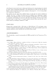
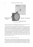
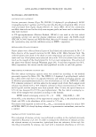
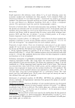
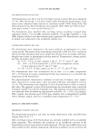
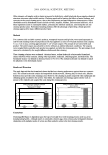
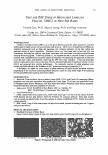
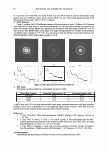
Micro-beam X-ray
(5 J1 m)
Diffraction
Hair fiber: 50 -.,100 J1 m
Figure 2. SAXS experiment.
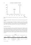
equal tilt to the hair axis. This diffraction pattern shows the same profile that was
estimated to diffract from the cuticle CMC in previous reports (6,7).
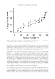
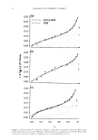
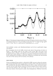
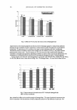
A one-dimensional intensity profile produced from the diffraction pattern is demon-
strated in Figure 3. The intensity {I(S) x S4} was plotted versus reciprocal spacing (S)
according to an analytical method proposed by Ohta et al. (7). Reciprocal spacing (S) was
used to represent the index for the distance from the center of the diffraction pattern,
where S =1/d =(2/A) x sin(20/2), with A representing the wavelength of the X-ray, 20
the scattering angle, and d the repeat distance. Using this method, we estimated the
thickness of the �- and 8-layers in the CMC. The region of S 0.2 shown in Figure 3
contains four peaks of I(S) x S4 .The spans of the peaks differed among the hair samples,
which reflected differences in CMC structure. All intensity profiles analyzed in the
present study contained at least three peaks of I(S) x S4 in the region of S 0.2. The
analytical region (S 0.2) was considered wide enough for correct estimation of the
thickness of the �- and 8-layers.
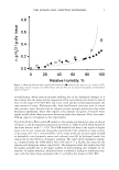
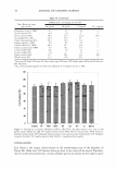
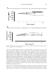
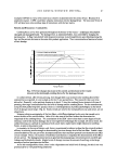
EFFECTS OF SOL VENT EXTRACTION ON CMC STRUCTURE
SAXS experiments were performed using hair samples extracted with the four solvents.
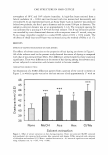
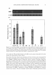
The estimated values of thickness of the �- and 8-layers differed, based on the solvent
used, as shown in Figure 4. The thickness of the �-layer was decreased by extraction with
acetone and hexane, while it was not changed by extraction with methanol or chloro-
form/methanol. In addition, the thickness of the 8-layer was decreased by extraction
Micro-beam X-ray
(5 J1 m)
Diffraction
Hair fiber: 50 -.,100 J1 m
Figure 2. SAXS experiment.
equal tilt to the hair axis. This diffraction pattern shows the same profile that was
estimated to diffract from the cuticle CMC in previous reports (6,7).
A one-dimensional intensity profile produced from the diffraction pattern is demon-
strated in Figure 3. The intensity {I(S) x S4} was plotted versus reciprocal spacing (S)
according to an analytical method proposed by Ohta et al. (7). Reciprocal spacing (S) was
used to represent the index for the distance from the center of the diffraction pattern,
where S =1/d =(2/A) x sin(20/2), with A representing the wavelength of the X-ray, 20
the scattering angle, and d the repeat distance. Using this method, we estimated the
thickness of the �- and 8-layers in the CMC. The region of S 0.2 shown in Figure 3
contains four peaks of I(S) x S4 .The spans of the peaks differed among the hair samples,
which reflected differences in CMC structure. All intensity profiles analyzed in the
present study contained at least three peaks of I(S) x S4 in the region of S 0.2. The
analytical region (S 0.2) was considered wide enough for correct estimation of the
thickness of the �- and 8-layers.
EFFECTS OF SOL VENT EXTRACTION ON CMC STRUCTURE
SAXS experiments were performed using hair samples extracted with the four solvents.
The estimated values of thickness of the �- and 8-layers differed, based on the solvent
used, as shown in Figure 4. The thickness of the �-layer was decreased by extraction with
acetone and hexane, while it was not changed by extraction with methanol or chloro-
form/methanol. In addition, the thickness of the 8-layer was decreased by extraction