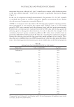
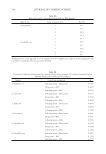
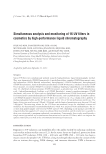
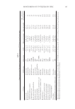
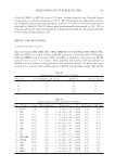
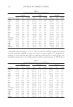
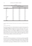
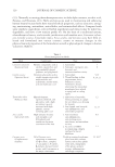
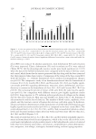
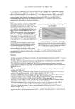
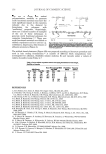
JOURNAL OF COSMETIC SCIENCE 136 Table I Effect of UVc Radiation Exposure on Minimum Inhibitory Concentration (MIC) on Staphylococcal Organisms Pen* MIC (μg/ml) Isolates UVc exposure time (min) Ery** MIC (μg/ml) Isolates UVc exposure time (min) 0 0.5 1.5 3 0 0.5 1.5 3 MRSA 3 ≥16 ≥16 ≥16 ≥16 MRSA 3 ≥32 ≥32 ≥32 ≥32 MRSA 20 ≥16 ≥16 ≥16 ≥16 MRSA 20 ≥32 ≥32 ≥32 ≥32 MRSA 80 ≥16 ≥16 ≥16 ≥16 MRSA 80 ≥32 ≥32 ≥32 ≥32 MRSA 143 ≥16 ≥16 ≥16 ≥16 MRSA 143 0.5 0.5 0.5 0.5 MSSA 63 0.03 0.03 0.03 0.03 MSSA 63 1 1 1 1 MSSA 93 0.03 0.03 0.03 0.03 MSSA 93 0.5 0.5 0.5 0.5 S. haemolyticus 16 ≥16 ≥16 ≥16 ≥16 S. haemolyticus 16 ≥32 ≥32 ≥32 ≥32 S. haemolyticus 17 ≥16 ≥16 ≥16 ≥16 S. haemolyticus 17 ≥32 ≥32 ≥32 ≥32 Cip*** MIC (μg/ml) Isolates UVc exposure time (min) Cip+res**** MIC (μg/ml) Isolates UVc exposure time (min) 0 0.5 1.5 3 0 0.5 1.5 3 MRSA 3 8 8 8 8 MRSA 3 8 8 8 8 MRSA 20 32 32 32 32 MRSA 20 32 32 32 32 MRSA 80 32 32 32 32 MRSA 80 32 32 16 16 MRSA 143 0.5 0.5 0.5 0.5 MRSA 143 0.25 0.25 0.25 0.25 MSSA 63 0.5 0.5 0.5 0.5 MSSA 63 0.25 0.25 0.25 0.25 MSSA 93 0.25 0.25 0.25 0.25 MSSA 93 0.25 0.25 0.25 0.25 S. haemolyticus 16 16 16 16 16 S. haemolyticus 16 8 8 8 8 S. haemolyticus 17 32 32 32 32 S. haemolyticus 17 32 32 32 32 MRSA = methicillin-resistant Staphylococcus aureus. MSSA = methicillin-sensitive Staphylococcus aureus. *Pen=penicillin **Ery=erythromycin ***Cip=ciprofl oxacin ****res=reserpine. particularly from within the clinical environment. Several environmental stresses induce the mar (multiple antibiotic resistance) operon (6), which regulates the expression of sev- eral genes, including those which encode a broad-specifi city effl ux pump (7). In addition, stress hardening (8) may lead to cross-protection against a range of apparently unrelated stress challenges, including resistance to antibiotics. Therefore, bacterial cells have sev- eral mechanisms to select for mutants from within sublethally stressed bacterial popula- tions, as well as to minimize stress and maximize continued cell viability to ensure survival following the removal of the stress conditions. More recently, the term “stresso- some” has been proposed, which describes a signal transduction cascade that increases the expression of stress-response genes and where stress signals may be integrated by a mul- tiprotein signalling hub that responds to various signals to effect a single outcome (9). In the current study, we wished to examine whether or not the sublethal stress relating to UV radiation of staphylococcal organisms led to an increase in the organism’s antibiotic resistance levels, by phenotypic examination of resistance to three common classes of an- tibiotics, namely the β-lactams (pencillin), the macrolides (erythromycin), and the fl uo- roquinolones (ciprofl oxacin). UV radiation exerts its bacteriocidal effect on bacterial populations through dimerization of adjacent thymine molecules on the organism’s

ANTIBIOTIC SUSCEPTIBILITY AND UVc LIGHT 137 DNA, thus leading to cell death. UVc light has a wavelength of between 280 and 200 nm and has the highest energy, compared to UVa and UVb light. Due to its absorption in the lithosphere, relatively little UVc light is experienced on the earth’s surface, in compari- son to UVa and UVb light, with the exception of high altitudes. Equally, cosmetic UV sunbeds do not emit UV light within the UVc range. Therefore, it would appear an usual choice of light on which to perform these experiments. UVc radiation was selected due to its highest energy and thus its ability to infl ict the greatest ability for bacterial mutational change and genomic re-organization, including alterations to the organism’s antibiotic susceptibility. Staphylococcal organisms were exposed to UVc radiation until the point of lethality, and their antibiotic susceptibility was examined along this time course. Very little is known about the effects of UV sunbeds on altering the diversity of the host bacterial commensal skin fl ora. Any UV radiation-induced alteration may led to a bacte- rial ecological imbalance, thereby leading to a vulnerability for the colonization and sub- sequent infection with skin pathogenic organisms. Overall, we were not able demonstrate any alteration in antibiotic resistance levels in any of the staphylococcal skin organisms examined, suggesting that UV stress response is not linked with upregulation of a global stress response cascade within the organisms examined. In conclusion, these in vitro data do not support any alteration in antibiotic susceptibility when challenged with sublethal stress from UVc light. ACKNOWLEDGMENT This work was fi nancially supported through an HSC R&D Offi ce commissioned grant: Antimicrobial Resistance Action Plan (AMRAP) (COM/2730/04). REFERENCES (1) D. E. Fisher and W. D. James, Indoor tanning—Science, behavior, and policy, New Eng. J. Med., 363, 901–903 (2010). (2) M. A. McMahon, J. Xu, J. E. Moore, I. S. Blair, and D. A. McDowell, Environmental stress and antibi- otic resistance in food-related pathogens, Appl. Environ. Microbiol., 73, 211–217 (2007). (3) P. Gilbert and A. J. McBain, Potential impact of increased use of biocides in consumer products on prevalence of antibiotic resistance, Clin. Microbiol. Rev., 16, 189–208 (2003). (4) A. P. Schuch and C. F. Menck, The genotoxic effects of DNA lesions induced by artifi cial UV-radiation and sunlight, J. Photochem. Photobiol. B, 99, 111–116 (2010). (5) Clinical and Laboratory Standards Institute (CLSI), Performance standards for antimicrobial susceptibil- ity testing. Document M100-S15 (Wayne, PA, 2005). (6) M. N. Alekshun and S. B. Levy, Alteration of the repressor activity of MarR, the negative regulator of the Escherichia coli marRAB locus, by multiple chemicals in vitro, J. Bacteriol., 181, 4669–4672 (1999). (7) A. H. Rickard, S. Lindsay, G. B. Lockwood, and P. Gilbert, Induction of the mar operon by miscella- neous groceries, J. Appl. Microbiol., 97, 1063–1068 (2004). (8) N. J. Rowan, Evidence that inimical food-preservation barriers alter microbial resistance, cell morphol- ogy and virulence, Trends Food Sci. Technol., 10, 261–270 (1999). (9) J. Marles-Wright, T. Grant, O. Delumeau, G. van Duinen, S. J. Firbank, P. J. Lewis, J. W. Murray, J. A. Newman, M. B. Quin, P. R. Race, A. Rohou, W. Tichelaar, M. van Heel, and R. J. Lewis, Molecular architecture of the “stressosome,” a signal integration and transduction hub, Science, 322(5898), 92–96 (2008).
Purchased for the exclusive use of nofirst nolast (unknown)
From: SCC Media Library & Resource Center (library.scconline.org)