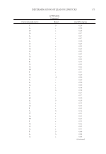
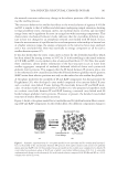
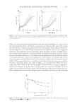
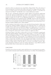
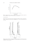
J. Cosmet. Sci., 63, 197–203 (May/June 2012) 197 Melanosome transfer evaluation by quantitative measurement of Pmel 17 in human normal melanocyte–keratinocyte co-cultures: Effect of an Alaria esculenta extract CLOTILDE VERDY, JEAN-ERIC BRANKA, and NICOLE MEKIDECHE, BIOTECHMARINE, Zone industrielle, BP 65, 22260 Pontrieux (N.M.), and EFFISCIENCE, 178 rue de Brest, 35000 Rennes (C.V., J.-E.B.), France. Accepted for publication November 8, 2011. Synopsis Numerous strategies have been proposed to evaluate melanosome transfer. Methods allowing quantitative measurements of this transfer in human normal cellular models, however, are very few and often require ex- tremely specialized devices that are expensive and diffi cult to use. As a part of the melanosome-specifi c membrane-bound glycoprotein, Pmel 17 is released from the melano- some membrane by ectodomain shedding. We reasoned, therefore, that it should be possible to evaluate melanosome transfer by quantifying this “soluble” Pmel 17. The Pmel 17 ELISA assay developed permits a detection of 10 to 1000 ng/ml of this glycoprotein in human normal melanocyte–keratinocyte co-culture media. As expected, niacinamide, a well-known melanosome transfer inhibitor, signifi cantly reduced the Pmel 17 quantities found in the culture media. This validated our experimental design. We then used our model to show that a whitening cosmetic active compound, i.e., an Alaria esculenta extract, can (at least in part) enable a signifi cant decrease in the melanosome transfer to produce a lightening effect without affecting melanin production. This research provides a simple and effi cient method to quantify melanosome transfer in a human normal co-culture model. It is a particularly useful tool with which to facilitate the development of new active whit- ening compounds. INTRODUCTION Quantifi cation of melanosome transfer is important in cosmetics for the development of new whitening compounds. Although several methodological approaches have already been proposed (for a review, see reference 1), they often require complicated and expensive devices that preclude rapid and effi cient screenings. Address all correspondence to Jean-Eric Branka at jebranka@effi science.fr.

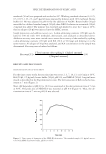
JOURNAL OF COSMETIC SCIENCE 198 In 2008, Singh et al. (2) proposed the silver locus product Silv/gp100/Pmel17 as a new tool for the analysis of melanosome transfer in human melanocyte–keratinocyte co-cultures. In this study, they demonstrated, in a human in vitro model, that double im- munofl uorescent labeling of keratinocytes and melanosomes permits the defi nition of Pmel 17 as a tracker of transferred melanin and of the melanosome transfer. This work could have provided a good base for our own work, but double immnofl uorescent labeling and the quantifi cation of the obtained signals did not fi t well with our goal of a “quick and simple” method to quantify melanosome transfer. We therefore decided to combine the methodological approach of Singh et al. with other data concerning Pmel 17. Proteolytic ectodomain shedding of Pmel 17 has been described to lead to the release of a soluble part in the extracellular medium (3,4). We therefore reasoned that it should be possible to evaluate melanosome transfer by quantifying this “soluble” Pmel 17. Antibodies that indifferently recognize the melanosome-bound and/or the soluble form of Pmel 17 are now commercially available, and so we chose to develop an ELISA assay to quan- tify Pmel 17 in the extracellular medium of in vitro melanocyte–keratinocyte co-cultures. We decided to evaluate the effect of niacinamide, a well-known inhibitor of melanosome transfer (5,6), in order to validate our experimental design. This is the focus of the second part of this study. Finally, and in order to show that our methodological and technological approach can be easily and effi ciently used to achieve screenings and/or later phases of the development of new whit- ening compounds, we examined whether the whitening effect of an Alaria esculenta extract currently used in cosmetics could be due to the reduction of melanosome transfer. MATERIALS AND METHODS REAGENTS AND MATERIALS Human normal keratinocytes were obtained from a 45-year-old Caucasian donor. Human normal melanocytes were obtained from a 35-year-old Caucasian donor. Keratinocyte growth medium 2 (KGM 2) and melanocyte growth medium 2 PMA free (MGM 2 PMA free) were purchased from Promocell (Heidelberg, Germany). Penicillin and streptomy- cin came from Invitrogen (Carlsbad, CA). Melanoma gp100 protein (Pmel 17) was pur- chased from Abcam (Paris, France). BSA fraction V and Tween 20 came from Acros Organics (Geel, Belgium). Human melanocyte protein Pmel 17 (SILV) antibody came from US Biologicals (Swampscott, MA). The secondary antibody, i.e., a goat anti-chicken IgY (H&L) horseradish peroxidase conjugated antibody, was purchased from Antibodies- online (Aachen, Germany). The substrate reagent for the peroxidase came from R&D Systems (Minneapolis, MN). H2SO4 came from Merck (Darmstadt, Germany). Multiwell culture plates, Transwell® inserts, and EIA/RIA Stripwell plates were purchased from Corning Costar (Brumath, France). The Alaria esculenta extract came from BIOTECH- MARINE (Pontrieux, France). CELL CULTURES AND TREATMENTS Normal human adult melanocytes were seeded in multiwell culture plates and cultured in MGM PMA free in a humidifi ed incubator under a 5% CO2/95% air atmosphere
Purchased for the exclusive use of nofirst nolast (unknown)
From: SCC Media Library & Resource Center (library.scconline.org)