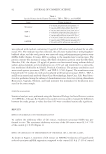
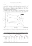
JOURNAL OF COSMETIC SCIENCE 148 IN VITRO MUSHROOM TYROSINASE INHIBITION TEST Enzyme activity was determined by the method described by Ishihara et al. (8), modifi ed according to the results of preliminary testing to search for optimized conditions. Each of the extracts and fractions was dissolved in 0.1 M sodium phosphate buffer (pH 6.5) and diluted to 1% individually. The solution was used as a test sample. Then, 20 μl of test solution, 220 μl of 0.1 M sodium phosphate buffer (pH 6.5), 40 μl of 1.5 mM tyrosine, and 20 μl of 2000 unit/ml mushroom tyrosinase were sequentially transferred to a 96-well plate and incubated at 37°C for 10 min. The absorbance was measured at 490 nm with an ELISA reader (GENios, Tecan, Männedorf, Switzerland). As a blank solution, 0.1 M sodium phosphate buffer (pH 6.5) was added, instead of test solution. IN VITRO INHIBITION OF L-DOPA AUTO-OXIDATION Enzyme activity was determined using the method described by Kong et al. (9), modifi ed according to the results of preliminary testing for optimized conditions. Each of the ex- tracts and fractions was dissolved in 0.1 M sodium phosphate buffer (pH 6.5) and diluted to 1% individually. The solution was used as a test sample. Then, 20 μl of test solution, 220 μl of 0.1 M sodium phosphate buffer (pH 6.5), 40 μl of 500 μM L -DOPA (Sigma- Aldrich Chemical Co., St. Louis, MO), and 20 μl of 2000 unit/ml mushroom tyrosinase were sequentially transferred to a 96-well plate and incubated at 37°C for 10 min. The absorbance was then measured at 490 nm with an ELISA reader. As a blank solution, 0.1 M sodium phosphate buffer (pH 6.5) was added instead of test solution. CELL CYTOTOXICITY TEST Neutral red assay. Transformed mouse fi broblast L929 was inoculated into a 96-well plate containing Dulbecco’s modifi ed Eagle’s medium (DMEM) with 2% bovine calf serum (BCS) (3 × 10³ cells/well) and incubated at 5% CO2 and 37°C for 48 h. The medium was then replaced with new serum-free medium containing transferrin (10 μg/ml) and ethanol- amine (2 μM) and again incubated for 48 h after the addition of sample dilutions. After incubation, 100 μl of neutral red (NR) solution (5 μg/ml) was added into each well and was allowed to react at 37°C for 3 h. After the reaction, the supernatant was removed and the solution was fi xed with 1.0% formalin containing 1.0% CaCl2 to immobilize cells for 5–10 min. After removal of the immobilization solution, 100 μl of 50% ethanol with 1.0% acetic acid was added to each well to extract pigments. The absorbance was then measured at 540–630 nm (double wavelength) with an ELISA reader to calculate the NR50. MTT assay. Transformed mouse fi broblast L929 was inoculated into a 96-well plate containing DMEM with 2% BCS (3 × 10³ cells/well) and incubated at 5% CO2 and 37°C for 48 h. The medium was then replaced with new serum-free medium contain- ing transferrin (10 μg/ml) and ethanolamine (2 μM) and again incubated for 48 h after the addition of sample dilutions. After incubation, 100 μl of 3-(4,5-dimethylthiazol- 2yl)-2,5-diphenyltetrazolium bromide (MTT) solution (5 mg/ml) was added to each well, and was allowed to react at 37°C for 4 h. After the reaction, the supernatant was removed and 100 μl of isopropanol containing 0.04 M HCl was added to each well to extract formazan from cells. The absorbance was then measured at 570–630 nm (double wavelength) with an ELISA reader to calculate the MTT50.

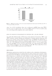
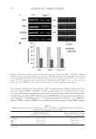
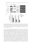
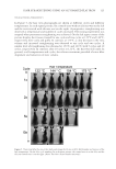
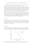
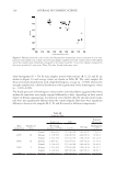
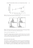
INHIBITORY EFFECTS OF KOREAN INDIGENOUS PLANTS 149 INTRACELLULAR TYROSINASE INHIBITION TEST From tyrosinase inhibition and cell cytotoxicity tests, four samples with a good inhibition effect and no cytotoxicity were selected to conduct intracellular tyrosinase inhibition and melanin synthesis tests. A six-well plate containing DMEM media (Gibco, Carlsbad, CA) with 10% fetal bovine serum (FBS Gibco) (2 × 10⁴ cells/well) was inoculated with 2 ml of murine melanoma (B16) cells and incubated at 5% CO2 and 37°C (CO2 air-jacketed incubator NuAire, Plymouth, MN) for 24 h. The medium was then replaced with fresh DMEM containing 10% FBS, 2 μM α-melanocyte-stimulating hormone (MSH), and 2 mM theophylline. Selected plant extracts and fractions were diluted with fresh medium and incubated with cells at 5% CO2 and 37°C until more than approximately 80%. After incubation, the medium was removed, cells were washed with phosphate-buffered saline (PBS), and trypsin was applied for the recovery of cells. The cell pellet was trans- ferred to an eppendorf tube and centrifuged at 10,000 rpm for 10 min to remove the supernatant. The cell pellet was washed two or three more times with PBS and centrifu- gation was repeated. Then, 100 μl of 0.1% Triton X-100 was added to dissolve cells and obtain intracellular tyrosinase extract, and 220 μl of 0.1 M phosphate buffer (pH 6.5), 40 μl of 1.5 mM tyrosine solution, and 20 μl of tyrosinase extract were added sequentially (10). After reacting at 37°C for 60 min, the absorbance was measured at 490 nm with an ELISA reader. For a blank test, only B16 melanoma cells were incubated without sample and the recovered tyrosinase extract was added. INTRACELLULAR MELANIN SYNTHESIS INHIBITION TEST A six-well plate containing DMEM with 10% FBS (2 × 10⁴ cells/well) was inocu- lated with 2 ml of murine melanoma (B16) and incubated at 5% CO2 and 37°C for 24 h. The medium was then replaced with fresh DMEM containing 10% FBS, 2 μM α-MSH, and 2 mM theophylline. Murine melanoma B16 cells were grown, incu- bated, and processed until a pellet was formed as described earlier. The cell pellet was dried at 60°C. Then, 100 μl of 1 N NaOH was added to the pellet to dissolve intracellular melanin. After dilution of melanin with PBS, the absorbance was measured at 490 nm with an ELISA reader to determine the melanin synthesis inhibition rate. For a blank test, only B16 melanoma cells were incubated without sample and the recovered tyrosinase extract was added. IN VIVO DEPIGMENTING EFFICACY TEST Based on the results of the in vitro tyrosinase inhibition test, in vitro L -DOPA auto- oxidation inhibition test, intracellular tyrosinase inhibition test, intracellular melanin synthesis inhibition test, and cell cytotoxicity test, the EtOAC fraction of P. lactifora and BuOH fraction of E. offi cinalis were proven to have good depigmenting effects and no toxicity, and were then subjected to an in vivo effi cacy study using guinea pigs to estimate the depigmenting effect in human body. Four female brown guinea pigs were used for an in vivo depigmentation study. To in- duce pigmentation, the backs of the animals were completely shaved with an electric
Purchased for the exclusive use of nofirst nolast (unknown)
From: SCC Media Library & Resource Center (library.scconline.org)