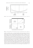
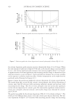
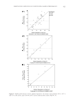
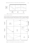
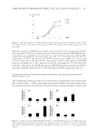
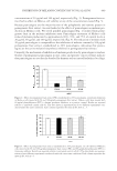
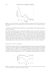
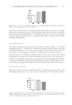
JOURNAL OF COSMETIC SCIENCE 432 fi ve per polycarbonate cage in a temperature (20°C) and humidity (40–45%)-controlled room. The light/dark cycle was 12:12 h and food (Samyang, Wonju, Korea) and water were supplied ad libitum. DETERMINATION OF HAIR GROWTH–PROMOTING ACTIVITY Hair growth–promoting activity of the L. esculentum extract was determined by the method reported by Roh et al. (25), with some modifi cations. Briefl y, 6-week-old C57BL/6 mice were randomly divided into fi ve groups for fi ve treatments, as follows: the negative control (NC) group (10% ethanol as a vehicle), positive control (PC) group (3% minoxidil), L. esculentum extract 1 group [3% (w/w) of EAE], extract 2 group [3% (w/w) of supercritical CO2 extract (SCE)], and extract 3 group [3% (w/w) of LTS]. Hair was removed from the 2 cm × 3 cm dorsal area of these mice by shaving carefully with an electric clipper. The substances and test materials were applied topically on the back skin of the mice, once a day for 4 weeks. The hair growth–promoting activity of the substances was checked by the darkening of the dorsal skin, which indicated the anagen phase of the hair follicles. The hair growth scoring was performed by two independent dermatologists, who were unaware of treatment regimen and we used the average of the two scores. Hair growth was measured at every 1 week during 4 weeks by assigning a hair growth index, as follows: score 0 = no growth observed 1 = up to 20% area of skin covered with hair 2 = 20–40% area of skin covered with hair 3 = 40–60% area of skin covered with hair 4 = 60–80% area of skin covered with hair and 5 = 80% to full area of skin covered with hair observed. Digital images of total hair growth on day 28 were obtained using Nikon Coolpix P100 (Nikon Co., Tokyo, Japan). RNA EXTRACTION AND REAL-TIME RT-PCR Total RNA extraction was performed with Trizol reagent (Life Technologies, Gaithers- burg, MD), and the cDNA was synthesized by a reverse transcription reaction using the RNA PCR kit (Applied Biosystems Roche Inc., Foster City, CA) in a 20 μl mixture containing 1 μg RNA, 50 mM KCl, 10 mM Tris/HCl, 5 mM MgCl2, 1 mM of each dNTPs, oligo-(dT) primers, 20 units of RNAse inhibitor, and 50 units of MuLV reverse transcriptase. Nucleotide sequence of the primers used in this study is shown in Table I. The reaction mixture was incubated for 60 min at 42 °C, then heated at 90°C for 7 min in a thermocycler (GeneAmp PCR system 9600 PerkinElmer, Roche Molecular System, Branchburg, NJ). Real-time PCR was performed using a lightcycler instrument using FastStart DNA Master SYBR Green PCR kit (Roche, Mannheim, Germany). Quantifi - cation of the VEGF, keratinocyte growth factor (KGF), IGF-I, and TGF-β mRNA ex- pression was corrected by glyceraldehyde 3-phosphate dehydrogenase (GAPDH). TEST HAIR TONIC WITH L. ESCULENTUM EXTRACT For possible future application in hair growth–promoting agents and pharmaceutical products for hair growth and alopecia prevention, the test hair tonic solution with LTS was prepared and evaluated for hair growth–promoting activity. At fi rst, 70 ml aliquots

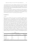
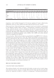
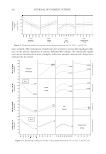
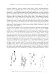
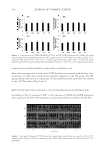
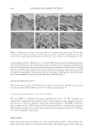
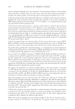
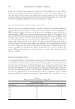
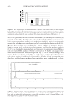
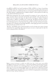
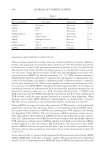
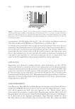
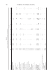
HAIR GROWTH–PROMOTING EFFECT OF L. ESCULENTUM EXTRACT 433 of distilled water were distributed into a glass beaker. A 0.3 g nicotine amide and 0.5 g salicylic acid were added and dissolved, and then the fi nal volume was brought up to 100 ml immediately. The formulations studied were prepared in a PRIMIX RM homomixer (PRIMIX Co., Ltd., Osaka, Japan) at 500 rpm within 5 min and supplemented with 3% (w/w) of LTS. A placebo formulation was prepared without LTS. HISTOLOGICAL ANALYSIS OF HAIR FOLLICLES The substances and test materials were applied topically on the back skin of the mice, once a day for 4 weeks. After week 4, all of the mice were sacrifi ced. Their dorsal skins were removed and fi xed in 4% formaldehyde solution and embedded in paraffi n. The fragments were sectioned into two different patterns: transverse sections for determina- tion of hair follicle count and longitudinal sections for the overall histological assessment. The 3-μm sections were stained with hematoxylin–eosin and toluidine blue and exam- ined under a light microscope (Magnifi cation: ×200) (Olympus, Melville, NY). DRAIZE SKIN IRRITATION TEST The irritation potentials of the test hair tonic solution with 3% LTS were evaluated accord- ing to the method of Draize (26) with slight modifi cation (27). Briefl y, two male New Zealand White (NZW) rabbits weighing 2.5–3.0 kg were acclimatized for 5 days before starting the study. The back of each rabbit was clipped free of hair and then divided into four sectors as shown in Fig. 8: after abrasion and application of hair tonic (upper left), after abrasion and no application of hair tonic (upper right), no abrasion and application of hair tonic (lower left), and no abrasion and no application of hair tonic as NC (lower right). The area of each sector was 6.25 cm2 (2.5 cm ×2.5 cm). For abrasion, several layers of skin were removed with adhesive tape from one half (upper side) of the shaved backs. Adhesive tape stripping was done about fi ve times. A 0.5 ml of the test hair tonic solution with 3% LTS was applied once, uniformly, on the left side only of the hair-free skin of each rabbit. Table I Nucleotide Sequence of the Primers Used For PCR Amplifi cations in This Study Growth Factor Primer Sequence VEGF Forward ACS CGG TGG TGG AAG AAG AG Reverse CAA GTC TCC TGG GGA CAG AA KGF Forward ACG AGG CAA AGT GAA AGG GA Reverse TGC CAC AAT TCC AAC TGC CA IGF-1 Forward TCA TGT CGT CTT CAC ACC TCT TCT Reverse CCA CAC ACG AAC TGA AGA GCA T TGF-β Forward GCG GCA GCT GTA CAT TGA CT Reverse ACT GTG TGT CCA GGC TCC AA GAPDH Forward CAA TGA ATA CGG CTA CAG CAA C Reverse AGG GAG ATG CTC AGT GTT GG
Purchased for the exclusive use of nofirst nolast (unknown)
From: SCC Media Library & Resource Center (library.scconline.org)