JOURNAL OF COSMETIC SCIENCE 380 Absorbance was read with a microplate reader (MultiskanMK3, Thermo Scientifi c, Waltham, MA) at 490 nm. Cell viability rate (%) of the samples against the proliferation of B16F10 was calculated using the following equation Cell viability rate (%) = Absorbance of well with samples/Absorbance of well without samples × 100 MELANIN ASSAY B16F10 cells (2 × 10⁵ cells) were cultured in DMEM with 10% FBS. After 12 h, the cells were treated with various concentrations of the samples or media only as a blank for 1 h. Following treatment, 100 nM α-MSH was added to the cells and incubated at 37°C with 5% CO2 in a humidified atmosphere. After 48 h, the cells were washed with phosphate- buffered saline (PBS) and harvested (5000 rpm × 10 min). The pellets containing a known number of cells were dissolved in 1 N NaOH solution containing 10% DMSO and sonicated for 1 h. The amount of melanin content was then monitored by a micro- plate reader at 490 nm. Data are expressed in terms of melanin synthesis inhibitory activ- ity compared to the control. Inhibitory activity (%) was calculated using the following equation. Inhibitory activity (%) = [1 − (Absorbance of sample − Absorbance of blank)/Absorbance of control] × 100. CELLULAR TYROSINASE ACTIVITY ASSAY Cellular tyrosinase activity was determined based on a modifi cation of a previously de- scribed method (8). As many as 2 × 10⁴ cells/cm2 were treated with the samples for 1 h, followed by addition of 100 nM α-MSH. After 48 h, the incubated cells were harvested and washed twice with ice-cold PBS by centrifugation at 1000 × g for 5 min. The har- vested cells were lysed in 1% Triton X-100 and 0.1 mM PMSF in PBS. The total protein was collected by centrifugation at 10,000 × g for 25 min at 4°C. The reaction mixture consisted of 100 μl 1 mg/ml L -DOPA solution and 50 μl cell-extracted protein. Dopach- rome formation at 37°C for 30 min was measuring the absorbance at a wavelength of 475 nm using a microplate reader. WESTERN BLOT ASSAY B16F10 cells were treated with the samples for 1 h, and α-MSH was added. After 48 h, cells were collected and lysed in a RIPA cell lysis buffer 3 containing 50 mM Tris-HCl, pH 7.4, 150 mM NaCl, 1 mM EDTA, 1 mM EGTA, 1.2% Triton X-100, 0.5% sodium deoxycholate, and 0.1% SDS (Enzo Life Sciences, Inc., Plymouth, PA). The lysates were denatured at 95°C for western blot assay. Proteins were separated using 10% SDS-poly- acrylamide gel electrophoresis running gel. The resolved proteins were transferred to ni- trocellulose membranes and then were blocked using 5% skim milk in Tris HCl buffer. Membranes were incubated with tyrosinase and GAPDH antibodies. And the membranes were washed three times every 15 min then incubated with anti-mouse horseradish

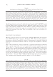
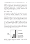
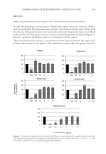
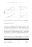
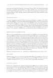
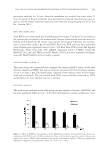
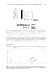
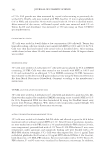
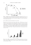
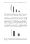
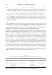
NELUMBO NUCIFERA AND INHIBITED TYROSINASE ACTIVITY AND MELANOGENESIS 381 peroxidase antibody for 30 min. After the membrane was washed four times every 15 min, the bends of bound antibodies were detected by enhanced chemiluminescence re- agents, and the images of protein expression were obtained using imaging system (Li-Cor Inc., Lincoln, NE). TRP-1, TRP-2 mRNA ASSAY Total RNA was isolated with the Trizol-Reagent (Invitrogen, Carlsbad, CA) according to the instructions provided by the manufacturer. Reverse transcription reactions were per- formed with SuperScript III reverse transcriptase (Invitrogen) following the manufac- turer’s instructions, using 2 μg of total RNA. Real-time PCR reactions were performed using Taqman gene expression assays by the 7500 Real Time PCR system (AB Applied Biosystems, Foster City, CA). The mRNA expression levels of TRP-1 (assay ID: Mm00453201_m1) and TRP2 (assay ID: Mm01225584_m1) were normalized to Hprt1 (assay ID: Mm00446968_m1) as loading controls. ANALYTICAL HPLC OF JKTM-12 The purity of the two compounds was confi rmed by analytical HPLC using a diode array detector. Analytical HPLC was run on a Supelco discovery C18 reverse phase column, 25 cm × 4.6 mm, 5 μm. The mobile phase consisted of two solvents, water (0.05% formic acid) and acetonitrile. The run started with 100% water and then increased to 100% acetonitrile in 35 min the fl ow rate was 1 ml/min. STATISTICAL ANALYSIS The results were analyzed statistically using one-way analysis of variance (ANOVA), and the least significant differences (p 0.05) were determined according to Duncan’s t-test. Figure 2. Effect of JKTM-12 on tyrosinase activity. Mushroom tyrosinase was incubated with various con- centrations of JKTM-12 and L -DOPA as substrates. Kojic acid was used as positive control. Data are pre- sented as the mean ± SEM of three individual experiments, performed in triplicate. Values are signifi cantly different by comparison with untreated control. *p 0.05, **p 0.001.
Purchased for the exclusive use of nofirst nolast (unknown)
From: SCC Media Library & Resource Center (library.scconline.org)