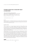
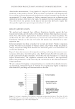
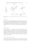
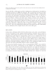
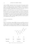
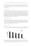
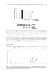
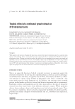
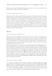
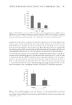
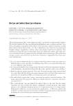
JOURNAL OF COSMETIC SCIENCE 378 oxidation of DOPA to dopaquinone, both of which comprise the fi rst stages of melanin synthesis (6). TRP-2 catalyzes the conversion of dopachrome to 5,6-dihydroxylindole-2 carboxylic acid (DHICA), and TRP-1 oxidizes the DHICA to indole-5,6-quinone car- boxylic acid and subsequently produces melanin (7). Therefore, not only tyrosinase in- hibitors but also TRP-1 or TRP2 inhibitors are of interest in the cosmetic industry for the treatment of hyperpigmentation and as skin-whitening agents (8,9). JKTM-12 is composed of the fl owers, roots, seeds, and receptacles of Nelumbo nucifera (Nymphaeaceae). N. nucifera, known as the sacred lotus, is used as food and medicine in East Asia and India (10). Various pharmacologically active substances, including alka- loids, fl avonoids, triterpenoids, polyphenols, steroids, and glycosides, have been extracted from different parts of N. nucifera (10). In this study, we investigated the inhibitory ef- fects of JKTM-12 on tyrosinase and melanin biosynthesis. Moreover, hyperoside and as- tragalin (Figure 1), which are the active compounds of JKTM-12 are shown to inhibit melanin biosynthesis, TRP-1 and TRP-2 mRNA expression, and cellular tyrosinase ac- tivity in B16F10 murine melanoma cells. MATERIALS AND METHODS MATERIALS Dulbecco’s modified Eagle’s medium (DMEM), fetal bovine serum (FBS), penicillin, and streptomycin were purchased from Invitrogen (Grand Island, NY). Glycerol tributyrate, L -tyrosine, mushroom tyrosinase, phenylmethanesulfonyl fl uoride (PMSF), kojic acid, and alpha-melanocyte-stimulating hormone (α-MSH) were purchased from Sigma- Aldrich (Steheim, United Kingdom). Monoclonal tyrosinase and GAPDH antibodies Figure 1. Chemical structure of astragalin and hyperoside.

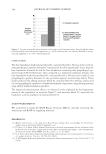
NELUMBO NUCIFERA AND INHIBITED TYROSINASE ACTIVITY AND MELANOGENESIS 379 were purchased from Cell Signaling Technology (Denver, MA). Anti-mouse horseradish peroxidase-conjugated immunoglobulin G (IgG) antibody was purchased from Santa Cruz Biotechnology (Santa Cruz, CA). ECL solution was purchased from Millipore Cor- poration (Billerica, MA). All other chemicals and solvents used in this study were of the analytical grade. PREPARATION OF JKTM-12 N. nucifera was harvested from the Muan-Gun area, South Korea, in August 2010 to Feb- ruary 2011. JKTM-12 (240 g) was composed of the fl owers (46 g), seeds (54 g), roots (60 g), and receptacles (80 g) of N. nucifera, respectively. JKTM-12 was prepared by boiling at 70°C with 70% EtOH for 3 h three times. It was then fi ltered and concentrated with a vacuum evaporator (N-3000, EYELA, Tokyo, Japan). After freeze-drying, the powder was stored at −4°C. The weight of the EtOH extract powder of JKTM-12 was 24.2 g (yield: 10.1%). JKTM-12 (EtOH extract powder) was dissolved in DMSO when used. MUSHROOM TYROSINASE INHIBITORY ASSAY The assay was performed using relevant methods (9). JKTM-12 each sample was dis- solved in DMSO to a fi nal concentration of 200 mg/ml. This extract stock solution was then diluted to 100 μg/ml in 100 mM potassium phosphate buffer (pH 6.8). Serial dilu- tions were made to attain fi ve concentrations. Kojic acid was used as a positive control. In a 96-well plate, 20 μl of each extract serial dilution was combined with 20 μl of mush- room tyrosinase (250 Units/ml in phosphate buffer) and 100 μl of 100 mM potassium phosphate buffer in triplicate. After incubation at room temperature for 5 min, 40 μl of the substrate (100 unit L -DOPA) was added to each well. The fi nal concentrations of the ex- tract samples ranged from 100 to 2000 μg/ml. After incubation at 37°C for 15 min, optical densities of the reaction mixtures in the wells were recorded at 490 nm using a BIO-TEK Power Wave XS multi-well plate reader (Bio-Teck Instruments, Winooski, VT). The activity rate was calculated by the following equation: Mushroom tyrosinase activity (%) = Absorbance of sample/Absorbance of control × 100. CELL CULTURE B16F10 murine melanoma cells were supplied by KCLB (Seoul, South Korea). They were cultured in DMEM supplemented with 10% heat-inactivated FBS, 10 U/ml of penicillin, 10 μg/ml of streptomycin in a 37°C, 5% CO2, 95% air humidifi ed atmosphere. ASSAY OF CELL VIABILITY B16F10 cell viability by the samples was measured by MTS assay according to the in- structions provided by the manufacturer. Briefl y, B16F10 cells were plated in 96-well culture plates (1 × 10⁴ cells per well). After 24 h, the media were changed and the cells were treated with the samples at various concentrations for 24 h. Ten microliters of MTS (5.0 μg/μl) was added to each well for an additional 4 h of incubation at 37°C.
Purchased for the exclusive use of nofirst nolast (unknown)
From: SCC Media Library & Resource Center (library.scconline.org)