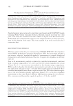
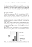
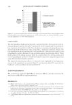
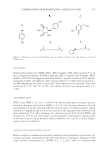
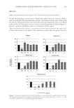
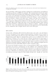
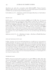
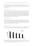
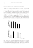
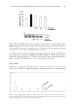
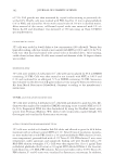
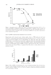
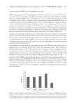
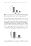
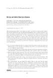
JOURNAL OF COSMETIC SCIENCE 384 fl owers, roots, and seeds of N. nucifera (data not shown). In our previous study, we isolated two bio-active compounds (hyperoside and astragalin) from an n-EtOAc fraction using the receptacles of N. nucifera. Hyperoside (IC50 = 15.67 μg/ml) and astragalin (IC50 = 21.22 μg/ml) were shown to inhibit tyrosinase activity (13). In this study, we demon- strated that hyperoside and astragalin inhibit melanine synthesis, TRP-1 mRNA, TRP-2 mRNA expression, cellular tyrosinase activity, and tyrosinase protein level. Hyperoside and astragalin were not cytotoxic to melanoma cells at the indicated concentrations (Fig- ure 4A). However, pretreatment of hyperoside and astragalin suppressed melanin biosyn- thesis overexpressed by α-MSH in B16F10 cells in a dose-dependent manner (Figure 4B). Because TRP-1 and TRP-2 are known as melanogenic enzymes, we investigated whether hyperoside and astragalin regulated TRP-1 and TRP-2 mRNA expression. As shown in Figure 5, TRP-1 and TRP-2 mRNA were overexpressed by 100 nM α-MSH in 48 h. However, pretreatment of hyperoside and astragalin inhibited TRP-1 and TRP-2 mRNA expression. Cellular tyrosinase and tyrosinase protein level were activated by 100 nM α-MSH, but pretreatment of hyperoside (10 μg/ml) and astragalin (10 μg/ml) inhibited Figure 5. Effects of hyperoside and astragalin on the mRNA expresstion of (A) TRP-1 and (B) TRP-2 in B16F10 cells. Cells were incubated in 6 well plates (2 × 10⁵ cells/well) with the indicated concentrations of hyperoside or astragalin for 1 h and then exposed to 100 nM α-MSH for 48 h. mRNA expression of TRP-1 and TRP-2 was detected by real-time PCR. Hprt1 was used as a loading control. Data are presented as the mean ± SEM of three individual experiments, performed in triplicate. *p 0.05 and **p 0.01 versus the only α-MSH-treated control values.

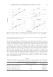
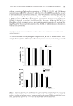
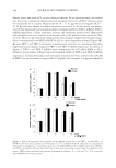
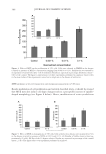
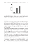
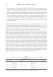
NELUMBO NUCIFERA AND INHIBITED TYROSINASE ACTIVITY AND MELANOGENESIS 385 the cellular tyrosinase activity and tyrosinase protein expression (Figure 6). Hyperoside and astragalin were identifi ed in JKTM-12 by HPLC (Figure 7). The retention times of hyperoside and astragalin were 20.519 and 22.902 min and the concentration of hypero- side and astragalin in JKTM-12 was 5.41 and 49.02 μg/g, respectively. DISCUSSION JKTM-12 is composed of the fl owers, roots, seeds, and receptacles of N. nucifera (the sacred lotus). JKTM-12 inhibited mushroom tyrosinase activity in a dose-dependent Figure 6. Effects of JKTM-12, hyperoside, and astragalin on the (A) cellular tyrosinase and (B) tyrosinase protein expression in B16F10 cells. Cells were incubated in 6-well plates (2 × 10⁵ cells/well) with JKTM-12 (JKTM, 100 μg/ml), hyperoside (Hyp., 10 μg/ml), or astragalin (Ast., 10 μg/ml) for 1 h and then exposed to 100 nM α-MSH for 48 h. Data are presented as the mean ± SEM of three individual experiments, performed in duplicate. *p 0.05 versus the only α-MSH treated control values. Figure 7. Identifi cation of hyperoside and astragalin in JKTM-12. Hyperoside and astragalin in JKTM-12 were identifi ed by HPCL following the protocols in the experimental section. The retention times of hypero- side and astragalin were 20.519 and 22.902 min, respectively.
Purchased for the exclusive use of nofirst nolast (unknown)
From: SCC Media Library & Resource Center (library.scconline.org)