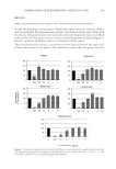
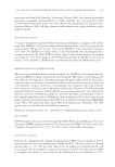
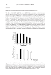
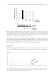
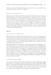
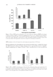
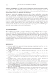
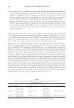
JOURNAL OF COSMETIC SCIENCE 398 Figure 8. Effect of MYE on actin cytoskeleton rearrangement. 3T3 cells were treated or not (control) with MYE at 0.1% during 90 min. (A) F-actin and G-actin were demonstrated by fl uorescence microscopy after labeling with Texas-red-phalloidin and DNAse I-FITC, respectively. The fi gure is representative of three independent experiments. (B) Densitometry analysis of F-actin-associated fl uorescence. For both control and MYE-treated conditions, 10 cells were sampled by using Image J software.

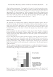
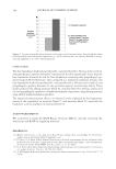
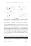
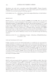
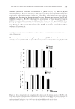
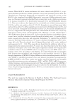
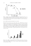
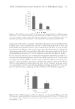
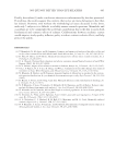
EFFECT OF METHANOL YEAST EXTRACT ON 3T3 FIBROBLAST CELLS 399 CONCLUSION Dermal connective tissue primarily comprises a matrix of hydrated protein fi bers such as collagen, elastin, fi bronectin, and glycosaminoglycans. This extracellular structure that is synthesized and colonized by fi broblasts serves both as mechanical support and exchange surface for the epidermis. Decrease of skin fi broblast activity caused by age and environ- mental stresses induces a dermal thickness reduction leading to the appearance of wrin- kles and a loss of elasticity. On the other hand, progress made in the fi eld of medical research, consistently increased our life expectancy often inducing a mismatch between health status and physical appearance. Hence, interest for cosmetics aimed to reduce time effect is still growing. In order to counteract skin aging, development of effective treatments able to sustain or increase fi broblast activity and proliferation is essential. In this regard, use of yeast ex- tracts appears to be a valuable and attractive approach. Indeed, despite the fact that it is a by-product of brewing industry, yeast is a natural and organic raw material rich in bio- active compounds. Research conducted by our group and others have highlighted the biological activity of a methanol extract able to stimulate S. cerevisiae metabolism and proliferation (16–19). Demonstration that this extract may also induce such a trophic effect on fi broblast cells will be a fi rst step for the formulation of a new topic antiaging treatment. Hence, in this study, we analyzed the effect of this MYE on various biological processes such as mitochondrial activity, proliferation, senescence, apoptosis, cytoskele- ton rearrangement, and migration of 3T3 fi broblast cells. Results showed that, up to a concentration of 0.1%, MYE is not cytotoxic and induces, after 72 h of incubation, a signifi cant increase of 3T3 cell proliferation. As suggested by clonogenicity experiments, the growth stimulation may be the result of both an increase of fi broblast number entering in cell cycle and a decrease of the generation time. Accord- ingly, we demonstrated that the yeast extract decreases both senescence and apoptosis when serum is depleted from culture medium (partially or totally, respectively). In Figure 9. Effect of MYE on the migration of 3T3 cells. After scratching cell monolayer with 2.2-mm-wide scrapper and treatment with mitomycin C, cells were incubated 24 h in the absence (control) or presence of MYE to 0.01% and 0.1%. Data represent the increase of cell migration after 24 h (i.e., decrease of the wound size). The fi gure is representative of four independent experiments performed in triplicate.
Purchased for the exclusive use of nofirst nolast (unknown)
From: SCC Media Library & Resource Center (library.scconline.org)