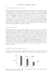
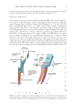
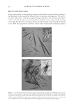
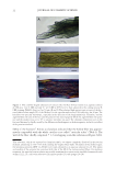
J. Cosmet. Sci., 66, 15–29 (January/February 2015) 15 New aspects of the structure of human hair on the basis of optical microscopic observations of disassembled hair parts ASAO YAMAUCHI and KIYOSHI YAMAUCHI, Department of Biochemistry, Osaka Municipal Technical Research Institute, Joto-ku, Osaka 536-8553 (A.Y.), and Keratin Materials Research Laboratory, Nishikyo-ku, Kyoto 610-1101(K.Y.), Japan. Accepted for publication November 23, 2014. Synopsis Infant’ and adult’ scalp hair fi bers were disassembled to various cellular components and blocks by chemical and enzymatic treatments, followed by random scission with rapidly rotating cutters. The hair fi bers were also fractured by the use of a vise. The optical microscopic inspection of these specimens led to the discovery of many previously unknown structures in the hair shaft. In particular, a cuticular cell (Cu) was found to take a trowel-like shape consisting of a part with a blade-like shape (CuB) and a part with a handle-like shape (CuH), where CuB overlapped one another and fused partially to build the honeycomb-like structure on a large cuticular thin plate (CuP). Whereas CuH was closely similar to the cortical cell in dimensions and rich- ness of macrofi brils (Mf). It was considered that human hair is stabilized structurally and physicochemically by the presence of the honeycomb-like structure, the CuP and the Mf. INTRODUCTION Several studies on the science of human hair were carried out over the last several decades, using the traditional structural model that was chiefl y composed of spindle-like cortical cells (Co), fl at cuticular cells (Cu), and serially aligned medulla cells (1–3). It would be very important, especially in a cosmetic fi eld, to analyze how chemical and biochemical agents interact with these cellular components. As a matter of fact, mammals’ hair fi bers have been frequently investigated by electron microscopes (4–15). The technique, however, even ignoring a laborious process for the preparation of a specimen, works in vacuo and provides only a black-and-white photograph of the dried substance. On the other hand, optical micro- scopes have been conveniently employed for examination of various hair samples despite the fact that the resolution is limited to semimicro and micrometer levels. Indeed, the micros- copy is well suited to dealing with wet substances, but also is very useful to inspect small objects for the structural characteristics, providing a see-through image with a high depth of fi eld and permitting chromatic distinction between two similar matters in the specimen. Address all Correspondence to Asao Yamauchi at yamauchi@omtri.or.jp
Purchased for the exclusive use of nofirst nolast (unknown)
From: SCC Media Library & Resource Center (library.scconline.org)