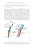
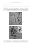
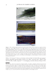
JOURNAL OF COSMETIC SCIENCE 16 The purpose of this study was to discover the unknown structures hidden in the inner domain of a human hair shaft. For achieving this aim, scalp hairs were disassembled or fractured to cellular components and blocks by combining chemical or enzymatic treatment with special cutting and compression processes. Taking advantages of the aforementioned optical microscopy, many specimens were analyzed for the inner structure of the hair shaft. EXPERIMENTAL HAIR SAMPLES AND REAGENTS Two- and six-year-old Japanese girls kindly gave black hairs I and II, respectively. Their hair was cut from more than 1 cm from the scalp surface. Virgin black hair III was simi- larly obtained from a healthy 16-year-old girl of the Miao ethnic group living in the mountains of China’s Yunnan province, and white hair IV from a 65-year-old Japanese male. All the hair fi bers were straight, neither being stained with dyes nor subjected to any permanent setting processes. The hairs were successively washed with aqueous 1.5 wt.% aqueous sodium dodecyl sulfate (SDS), deionised water, and aqueous 70 vol.% ethanol then the hairs were stored at 4°C in a sealed plastic container. All reagents, in- cluding 2-mercaptoethanol (ME), staining dyes, and papain, were commercially available. PREPARATION OF THE SPECIMENS FOR OPTICAL MICROSCOPY† The following four methods were adapted to prepare the specimens for microscopy. Meth- ods 1–3 consisted of the chemical and enzymatic pretreatments of the hair fi bers and the special cutting processes. Method 4 utilized a mini-vise to fracture the hair shaft. Method 1: Simple swelling treatment of hair fi bers by heating in the presence of urea and SDS (a general procedure). Hair (I–IV, 20 mg, about 1 cm length) was kept in the aqueous solu- tion (5 ml) of 8 M urea and 4 wt.% SDS at 55°C for 5 h without stirring. Since the treat- ment solution did not contain ME, the hair shaft becomes soft without disturbance of the structure see the swelling degree.‡ The swollen shaft was washed briefl y with pure water, dispersed in water (1 ml), and subjected to the cutting process I (vide infra). The resulting †The benefi ts of staining of the dyes employed in the present study are as follows: CBB—The Mf of the Co and the Cu were nonspecifi cally stained blue. Congo red—In contrast with the Co, the Cu, particularly the blade-like shaped parts (CuB), were stained preferentially pale red. Gentian violet—This dye was milder than CBB, staining the cellular components in various depth of blue-violet. Giemsa—The Co, the handle-like shaped parts (CuH) of the Cu and the medulla’s inner substance were more intensely stained than CuB and the medulla’s wall. Silver—Almost of all proteinous substances were stained black. Silver staining was useful to recognize the presence of CuB that was hardly stainable with any of the organic dyes. SM—This dye has an Hg2+ ion in the molecule. Therefore, sulfur-rich cellular components or sites are preferentially stained in red (the color of the ligand) presumably due to a formation of a strong Hg–S linkage. ‡When hair (I–IV about 20 mm length in plain water at 25°C) was heated in an aqueous mixture of 7 M urea, 3.5 wt.% SDS and 0 or 15 wt.% ME at 80°C for 20 min–1 h, the shafts were swollen in the follow- ing degree which was estimated using the equation, 100 × (L2 – L1)/L1, where L1 and L2 are the breadth and length of the hair shaft before and after the treatment, respectively. On heating for 1 h in the absence of ME: (breadth) I 34 ± 6%, II 28 ± 4%, III 25 ± 7%, and IV 21 ± 8% (length) I~IV about 0%. On heating for 20 min in the presence of ME: (breadth) I 143 ± 7%, II 150 ± 15%, III 150 ± 13%, and IV 200 ± 20% (length) I 13 ± 4%, II 21 ± 2%, III 35 ± 2%, and IV 43 ± 3%. All the hair samples swelled maximally after heating for about 1 h: (breadth) I~IV 200 ± 15% (length) I~II 40 ± 3%, and III~IV 50 ± 10%.

NEW ASPECTS OF THE STRUCTURE OF HUMAN HAIR 17 aqueous suspension of small hair fragments was mixed with a fresh 0.5 wt.% aqueous dye solution (2–3 drops) at ambient temperature for 0.5–5 h, where the dye used was Congo red, Coomassie brilliant blue G-250 (CBB), Gentian violet, 2’,7’-dibromo-4’- hydroxymercurifl uorescein sodium salt (SM), and a Giemsa’s solution (Wako Pure Chem., Osaka, Japan) (16). Sometimes the hair fragments were silver stained by soaking in aque- ous 0.2 wt.% silver nitrate, separating with a centrifuge, and exposing to bright sunlight for a while, followed by reduction with an aqueous 2 wt.% ascorbic acid at ambient temperature. After washing briefl y with water, the stained sample was placed on a micro- scope glass slide using aqueous 50 wt.% glycerol as a mounting medium and overlaid with a cover glass (Matsunami Glass No.1). A weight was placed on the cover glass (15–30 g/cm2) while sealing the glass edges with Canada balsam. Method 2: Treatment of hair fi bers with papain (a general procedure). Hair (I–IV, 0.13 g, about 1 cm in length) was incubated with occasional shaking at 55°C for 2–6 h in the pH 7/0.07 M phosphate solution (10 ml) containing papain (crude powder type from Carica papaya, about 0.15 unit), L-cysteine (0.1 g, an activator), and SDS (0.23 g). The digested fi bers were taken out with a spatula at 2-h intervals. After washing with water, the fi bers were suspended in pure water (2.5 ml) and subjected to the cutting process I or III (vide infra). The resulting hair fragments were stained and mounted to a microscope glass slide in a manner similar to that mentioned above. Method 3: Chemical treatment of hair fi bers using ME (a general procedure). Hair (I–IV, 80 mg, about 2 cm in length) was put to an aqueous mixture (10 ml) of 6.4–8 M urea, 3–4 wt.% SDS and 1–20 wt.% ME in a screw-cap glass test tube. The tube was set in a horizontal position and warmed without shaking at 50°C–85°C for 20 min–12 h. After cooling to ambient temperature, the solid remainder of the hair shaft was washed briefl y with aque- ous 0.5 wt.% ME and subjected to the cutting process II or III (vide infra). The resulting suspension of hair fragments was centrifuged at 750 g to give the precipitate that was washed with aqueous 0.2 wt.% ME, stained, and mounted on the slide glass in a manner similar to that described above. Method 4: Compression fracturing of the hair shaft. Hair (III, IV) was warmed in an aqueous solution of 7 M urea and 3.5 wt.% SDS at 55°C for 5 h. The resulting softened fi ber was washed briefl y with water, cut into about 10 mm in length, and 2 or 3 pieces were sand- wiched between a glass slide (thickness, 1.3 mm) and a cover glass (20 × 20 mm with a thickness 0.72 mm) using Canada balsam as a medium. Subsequently, the glass plates were pressed by a mini-vise at the force which was slowly increased to about 0.5–1.0 kgf, taking about 15 min. The vise with the glass plates was then stored in a refrigerator (about 10°C) in order to harden the medium, followed by taking off the vise at ambient temperature to give the specimen for microscopic observation. By this compression method, the cuticular covering of the hair shaft was usually fractured in the same direc- tion as the fi ber’s longitudinal axis. MECHANICAL CUTTING PROCESSES (I, II, AND III) The chemically and enzymatically pretreated hair fi bers (obtained in the aforementioned methods 1–3) were randomly chopped using the following three kinds of cutters. (i) A stainless steel gear (18 teeth, 8 mm diameter and 4 mm height) was placed in the aqueous suspension of the pretreated hair fi bers at ambient temperature and rotated by a
Purchased for the exclusive use of nofirst nolast (unknown)
From: SCC Media Library & Resource Center (library.scconline.org)