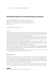
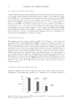
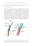
JOURNAL OF COSMETIC SCIENCE 18 small electric motor in the speed of 3500–4000 rpm for 3–10 s. This cutting method was found to effectively separate the blade-shaped parts (so-called “scale”) from Cu. (ii) A soft polyethylene gear having four teeth (each, 4 mm length × 2 mm width × 0.5 mm thickness) was rotated in an aqueous suspension of the pretreated hair fi bers in water (3–5 fi ber/ml) at the speed of about 500 rpm and ambient temperature for about 10 sec. This cutting process was used for the chemically or enzymatically pretreated hair fi bers to coarsely chop the shaft. (iii) A nose hair cutter (Hitachi, model BM-03, Tokyo, Japan) was used to obtain the very small hair fragments including the cells and cellular blocks. The outer steel blade with 9 slits (each slit: 0.8 mm width and 3.5 mm height) was designed to guide hair into a pair of inner blades (9 mm edge length, 2 mm depth, and 0.1 mm thickness). The pretreated hair fi bers in distilled water (1.5–2.0 cm3) were shortened with scissors to 1–5 mm length and put into the transparent plastic parabolic container (22 mm base diameter and 32 mm height), which was originally designed as the cap of the cutter head. The cutter body was Diagram 1. Schematic representation of the structures of a human hair shaft, the cortical cell (Co) and the cuticular cell (Cu). Co takes a spindle-like shape. The cells gather together to form more than 20 thick cord- like shaped substances. Cu is a trowel-like shaped substance, consisting of a handle-like-shaped part (CuH) and a CuB. CuH is similar in dimensions to Co, and both are fi lled with plenty of macrofi brils (Mf). Cu overlap one another and fuse partially, displaying tile roof-like and honeycomb-like patterns in the outer and inner surfaces of the CuB region, respectively. CuB, in its basal area, merges completely with other units to produce the CuP, which encircles the inner cellular components. The nucleus (N) of Cu is in CuB. Medulla (M) is a tubular substance in the center of the hair fi ber. The size (micrometer in unit) of the cells and the cellular components was measured by the use of an optical microscope and not corrected with the swelling degree of a hair shaft.‡

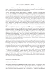
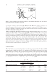
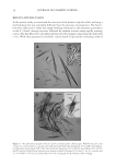
NEW ASPECTS OF THE STRUCTURE OF HUMAN HAIR 19 vertically clamped upside down, the container having the aqueous suspension of the fi bers was set to the cutter head, and then the inner blade was rotated for 5–7 s. MICROSCOPIC OBSERVATIONS The biological microscopes, Olympus Photomax LB and BHS with proper attachments, were used for bright fi eld, phase contrast, and polarized light observations. Objective lenses (Olympus) were as follows: (bright fi eld) DPlanApo 10×, 20×, and 40× CPL 20×/1.2 and LWDC Plan 40×/0.3–1.3. (Phase contrast) PL10×, PL20×, and PLL40× (polarized) P20× and P40× cf. the PlanApo lenses were usually used unless otherwise noted. Original photo tube accessories were modifi ed to adapt the digital camera that was automatically controlled by a desktop computer to optimize for lighting, ISO levels (400-6400), and focusing measures. The images in JPEG and RAW formats were devel- oped by means of Lightroom ver.3 and Photoshop Elements ver. 9 software (Adobe Systems Inc. San Jose, CA). Image enhancement included color level correction, noise reduction, and contrast and brightness adjustments the purpose of the enhancement was to make the image appear nearly identical to that seen actually by the observer. In the case of thick specimens, the images taken at various depths of fi eld were merged into a deep focus picture using the stacking software, CombineZM (A. Hadley, Sheffi eld, UK), (17). Diagram 2. Schematic representation of the side views of the Cu in a hair shaft. The cells overlap one an- other to form the honeycomb-like structure and the CuP. On the mechanical agitation, the cell is broken into the blade-like shaped part (CuB), the handle-like shaped part (CuH), and the fragmentary substance (CuB’).
Purchased for the exclusive use of nofirst nolast (unknown)
From: SCC Media Library & Resource Center (library.scconline.org)