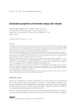
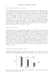
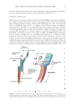
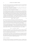
JOURNAL OF COSMETIC SCIENCE 20 RESULTS AND DISCUSSION In the present study, we examined the structure of the human scalp hair shaft, utilizing a methodology that was somewhat different from the previous investigations. The hair fi - bers were subjected to either the simple swelling treatment or the enzymatic proteolysis or the S–S bond cleavage reaction, followed by random scission using rapidly rotating cutters. The hair fi bers were also physically fractured by strongly compressing the shaft with a vise. With these preparative methods, various kinds of specimens containing isolated Figure 1. The photomicrographs of the Co and Cu including the cellular parts, PLL40× objective bar 25 μm. Co, cortical cell Cu, cuticular cell CuB, the broad blade-like shaped part of Cu CuB’, the fragmen- tary substance derived from CuB CuH, the handle-like shaped part of Cu Mf, the macrofi brils of Co and Cu and N, nucleus. Hair III was warmed in an aqueous solution of papain (0.15 unit), 1 wt.% L-cysteine and 2.3 wt.% SDS at 55°C for 3 h, then subjected to the cutting process II Gentian violet staining.

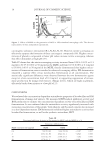
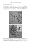
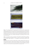
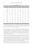
NEW ASPECTS OF THE STRUCTURE OF HUMAN HAIR 21 cells, cells’ blocks, fractured hair shaft, etc. were obtained. Like playing a jigsaw puzzle, the microphotographs of these disassembled hair parts were logically combined, resulting in the discovery of many unknown structures of the cellular components and the hair shaft itself. Some of them are schematically illustrated in Diagrams 1 and 2. The new structural features were common to all the hair samples (I–IV) examined. CORTICAL CELL The cell was easily recognized by a well-known spindle-like shape (a length of 80–90 μm and a width of 6–9 μm) as shown in Figure 1(A). The fi brous substances, which are most likely referred to as macrofi brils (Mf), were aligned in the same direction as the cell’s longitudinal axis. Though the Cos, together with a medulla, have long been considered to occupy the inner domain of a hair shaft (1–3,12) and affect the fi ber curvature (18–20), the cells were found to localize in the small space of the hair shaft, concentrically sur- rounding the medulla (M) (Figure 2). Furthermore, all the Cos appear to be grouped into more than 20 thick cord-like shaped substances [Figure 3(A)]. The substances twisted together as seen clearly in the specimens, which were prepared by physically fracturing the hair shaft [Figure 3(B) and 3(C)]. Formation of the cord-like shaped substances has not been explained well at present, but might be due to a special spatial arrangement of the Co in other words, every Co was regularly shifted a little from neighbors along the long and short axes of the cell body (Figure 4). By the way, the Co was often more deeply stained with the basic dye, Gentian violet, than the Cu the former cells were bluish purple while the latter, particularly the CuB part (vide infra), were gray or pale blue as shown in Figure 5. The facile stainability of the Co may be attributed to the high content of acidic amino acid residues of the keratin proteins involved (5,8,21). CUTICU LAR CELL On chemical treatment with an aqueous mixture of urea, SDS, and ME, the hair shaft was transformed to a hollow fi ber, gradually losing the inner substances [Figure 6(A) and Figure 2. The cross section of a hair shaft (left: no staining right: Congo red staining) bar 50 μm. The re- gion of the handle-like shaped part (CuH) of the cuticular cells (Cu) was more deeply stained by the dye than the region of the cortical cells (Co). Hair II was heated in an aqueous solution of 7 M urea, 3.5 wt.% SDS and 15 wt.% ME at 55°C for 15–30 min, followed by slicing with the cutting process III. Thereafter, about half the amount of the resulting substances was subjected to Congo red staining.
Purchased for the exclusive use of nofirst nolast (unknown)
From: SCC Media Library & Resource Center (library.scconline.org)