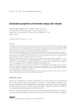
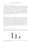
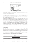
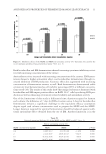
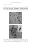
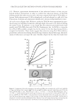
JOURNAL OF COSMETIC SCIENCE 22 6(B)] cf. the footnote.§ Amino acid analysis indicated that the hollow fi ber was approxi- mately comparable with the whole cuticles or so-called “cuticular scales” (Table I). The wall of the fi ber chiefl y comprised 7–12 overlapping scale-like substances [Figure 6(C)]. Figure 3. The cord-like shaped substances of cortical cells. (A) Hair III was treated in an aqueous solution of 8 M urea, 4 wt.% SDS and only 0.7 wt.% ME at 85°C for 2 h, then subjected to the cutting process II CBB staining PLL40× objective bar 50 μm. (B and C) The polarized light microscopic pictures of the frac- tured shaft of a hair sample III, cf. the Method 4 in the experimental section P40× objective bar 50 μm. The hair fi ber was optically anisotropic, especially in the direction of the longitudinal axis. Therefore, and the angle between the axis of the hair and the polarizer was varied properly while the angle between the polar- izer and the analyzer was set to 45° a sensitive tint plate was used. The chromatic distinction seen in the fractured hair may be chiefl y caused by the difference in birefringence or the heterogeneity in the α-crystallites of the proteins involved. §The substance, which was squeezed out from hair shafts, was almost completely dissolved in the reaction medium, amounting to 45%–70% of the starting dry weight of hair shafts. On dialysis of the mother liquor, the keratinous proteins (MW 12–60 kD) were easily obtained as an aqueous solution (21,22). The amino acid analysis of the proteins was consistent with that of the Mf of the starting natural fi bers. On oxidation treatment, the aqueous keratin solution was readily transformed to various water-insoluble biomaterials such as fi lm (21,22,23), cultivation substrata (24), microcapsules (25), and sponge (26–28).

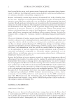
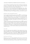
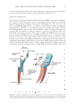
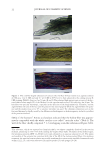
NEW ASPECTS OF THE STRUCTURE OF HUMAN HAIR 23 The high tolerance of the wall to the severe chemical treatment may be attributed to the proteins that were heavily cross-linked with the isopeptide [ε-γ(glutamyl)lysine] and disulfi de bonds (3–6 and 15–20 bonds per 1000 amino acid residues, respectively) (29, 30). On the other hand, the Cu was found to take the trowel-like shape, consisting of a trans- parent CuB and a CuH Figure 1(B) and the inset, cf. Diagram 1 (position: 10-m). The spatial arrangement of CuB and CuH were judged in Figure 7, i.e., CuH was attached to the outer surface of CuB cf. the schematic illustration in Diagram 2 (position: 6-b and 5-i). The cell itself, however, was not so stable on the mechanical impact given in the Figure 4. (A) A block of the cortical cells PL20× objective bars 25 μm. Hair I was heated in an aqueous mixture of 6.4 M urea, 3.2 wt.% SDS and 20 wt.% ME at 80°C for 7 h, followed by subjecting to the cutting process II CBB-staining. (B) Plenty of the fi brous substance in the cortical cells’ block bar 50 μm. The specimen was prepared by treating hair III in a manner similar to that mentioned in panel A, except for Gentian violet staining. Figure 5. (A) Cortical cell (Co), the blade-like shaped (CuB), and handle-like shaped (CuH) parts of the cuticular cell bar 50 μm cf. Figure 1. (B) CuB overlapped the other to display a tile roof-like or so-called “scale” pattern bar 50 μm. Every CuB possessed a nucleus (N) with 3–7 small black nucleoli. The specimens of panels A and B were obtained by heating hair (III) in a solution of 8 M urea and 4 wt.% SDS (without using ME) at 85°C for 15 h, followed by subjecting to the cutting process I Gentian violet staining.
Purchased for the exclusive use of nofirst nolast (unknown)
From: SCC Media Library & Resource Center (library.scconline.org)