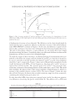
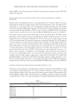
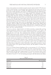
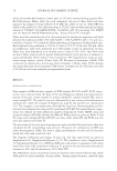
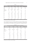
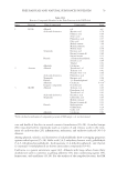
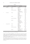
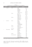
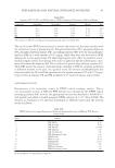
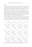
JOURNAL OF COSMETIC SCIENCE 86 The kinetic of the process was analyzed for each concentration tested, and the percentage of DPPH remaining at the steady state was estimated. This value was used to calculate the IC50 (defi ned as the concentration of substrate mg/ml that causes 50% loss of DPPH activity). Results are reported in Table XIV. The polar fractions of DM and WM showed a similar antioxidant activity (Table XIV, entries 1–3 vs. 7–9), higher than that observed for the corresponding nonpolar fractions (Table XIV, entries 1–3 vs. 4–6). In particular, EtOAc was the most active fraction in DM (IC50 2.0 × 10⁻³ mg/ml), and MeOH in WM (IC50 3.0 × 10⁻³ mg/ml). Evaluation of cytotoxic potential. The cytotoxic potential was evaluated in CHO cells only for the polar fractions, using the clonogenic assay (CA). CA enables an assessment of the differences in reproductive viability (capacity of cells to produce progeny, i.e., a single cell to form a colony of 50 or more cells) between control untreated cells and cells after treat- ment (69). Normally, only a hundred or few hundred cells are inoculated. Each viable cell grows and forms a colony. After a suitable incubation time (5–8 days), colonies are stained and counted manually. Cloning effi ciency is calculated as percentage (%) of colonies from all inoculated cells. Cytotoxicity was determined by measuring the RCE after treatment compared with cloning effi ciency of solvent control cultures. The assay was performed using a set of at least six dose levels for each extract, spaced by a factor of 2.0. DMSO was used as solvent. Results obtained indicated that after a 3-h treatment, dose level of 32.0 μg/ml proved to be toxic (RCE 20%) for all tested extracts, with the exception of WM-MeOH and DM-MeOH for which toxic levels were reached at 562.0 and 128.0 μg/ml, respectively (Tables XV–XX). Genotoxic and antioxidant activity in cultured mammalian cells. The antioxidant activity of polar and nonpolar fractions of DM and WM peloids was further evaluated in Table XV Analysis of Colony-Forming Activity for WM-Acetone Test compound Dose level (μg/ml) Mean of colonies RCE DMSO 1 317 100 WM-Acetone 1.0 311 98 2.0 310 98 4.0 276 87 8.0 200 63 16.0 171 54 32.0 10 3 Table XVI Analysis of Colony-Forming Activity for DM-Acetone Test compound Dose level (μg/ml) Mean of colonies RCE DMSO 1 298 100 DM-Acetone 1.0 292 98 2.0 265 89 4.0 224 75 8.0 143 48 16.0 54 18 32.0 12 4

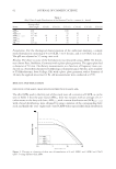
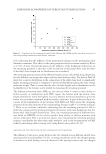
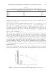
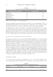
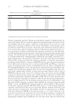
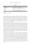
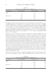
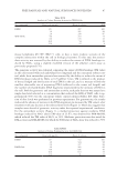
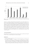
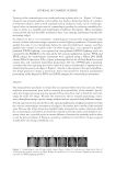
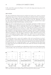
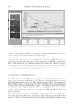
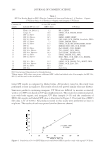
FREE RADICALS AND NATURAL SUBSTANCES IN PELOIDS 87 mouse lymphoma L5178Y (TK+/−) cells, to have a more realistic scenario of the complex interaction within the cell in biological systems. To this end, the antioxi- dant activity was assessed by the ability to reduce the extent of DNA breakage in- duced by H2O2, using a slightly modifi ed version of the alkaline comet assay as previously proposed (70). The genotoxic activity was evaluated comparing the extent of DNA breakage (TM value) in the cells treated with each individual test compound and the concurrent solvent con- trol, while their antioxidant potential was assessed by the ability to reduce the extent of DNA breakage induced by H2O2 at 0.25 μM for 5 min. TM is defi ned as the product of the tail length and the fraction of total DNA in the tail, and is a measure of both the smallest detectable size of migrating DNA (refl ected in the comet tail length) and the number of relaxed/broken DNA fragments (represented by the intensity of DNA in the tail). Both for genotoxic and antioxidant activity, each polar fraction was assayed at a single dose level selected as a concentration that reduced the RCE of CHO cells to ap- proximately 50% over the concurrent vehicle control cultures (Tables XV–XX). Selec- tion of dose levels was performed in previous experiments. For genotoxicity, the results indicated the absence of increase in the DNA migration (as measured by TM values), after treatment with any fraction at the selected dose levels (Figure 1). These data suggest that samples were devoid of genotoxic activity under the reported experimental conditions. For the antioxidant activity (Figure 1), a marked protection against oxidative DNA breakage induced by H2O2 alone (TM value of 23.07), was observed for WM-MeOH, which reduced the TM value of H2O2 to 34%. Moderate protection was also noted for DM-acetone and DM-MeOH, for which the TM value of H2O2 alone was reduced to 79% Table XVII Analysis of Colony-Forming Activity for WM-EtOAc Test compound Dose level (μg/ml) Mean of colonies RCE DMSO 1% 287 100 WM-EtOAc 1.0 281 98 2.0 250 87 4.0 215 75 8.0 135 45 16.0 54 18 32.0 12 4 Table XVIII Analysis of Colony-Forming Activity for DM-EtOAc Test compound Dose level (μg/ml) Mean of colonies RCE DMSO 1% 332 100 DM-EtOAc 1.0 324 98 2.0 327 98 4.0 282 85 8.0 186 56 16.0 110 35 32.0 41 12
Purchased for the exclusive use of nofirst nolast (unknown)
From: SCC Media Library & Resource Center (library.scconline.org)