EVALUATION OF MOLECULES OR EXTRACTS MODULATING SEBORRHEA 185 (100 U/ml penicillin, and 100 mg/ml streptomycin), 50 μg/ml EGF, and FCS. Mycoplasma removal treatment was achieved using Plasmocure® kit (Invivogen®, San Diego, CA) and followed regularly (MycoAlert® mycoplasma detection kit, Lonza®, Basel, Switzer- land). For lipid detection, proliferative NHSs were seeded in 96-well fl at and black bot- tom plate (Becton®, Rungis, France). When the cells reached confl uence, contact between cells and compounds was performed for 24–72 h. Cell number and lipid synthesis were estimated, respectively, using fl uorescein diacetate (FDA) and Red Nile fl uorescent probes (both from Sigma®-Aldrich®, St. Louis, MO) (8). Results were obtained using a fl uores- cent reader (FLUOstar®, BMGLabteck®, Ortenberg, Germany) equipped with appropri- ate fi lters. For lipid in situ labelling, cells were fi xed with Histochoice® (Clinisciences®, Nanterre, France) then incubated with Oil Red O® (Sigma®) solution and observed under microscope. For protein in situ labelling, cells were fi xed with Histochoice® then permeabi- lized with 0.1% Triton® X-100 (Sigma-Aldrich®) in phosphate-buffered saline and blocked in 1% bovine serum albumin/0.05% Tween® 20 (Sigma-Aldrich®). Primary antibodies were as follows: anti-K7, anti-K4, anti-fi laggrine, anti-involucrine, and anti-PLIN-2 (Santa Cruz Biotechnology®, Eurogentec®, Dallas, TX) (1). Species-specifi c secondary antibodies were conjugated to Alexa-Fluor® 488 (Life Technologies®, Liège, Belgium). Nuclei were counterstained with propidium iodide or Hoechst dye (Sigma-Aldrich®, Carlsbad, CA). For pro-infl ammatory studies, cells received for 24 h both Escherichia coli LPS (Aldrich®) and CCSV (IRB®, Altavilla Vicentina, Italy) for 3 days. Release of PGE-2 was evaluated using EIA assay (Cayman®). AntiMicrobial Peptide (AMP) cathelicidin and beta-defensin-2 (hBD2) were measured after 3 days from cell lysates using ELISA assays (Hycult Biotech®, Peprotech®, Peprotech, Rocky Hill, NJ). Results were normalized with cells quantifi ca- tion through Hoescht 33258 (Uden, The Netherlands) method. Collagen lattices were pre- pared according to the method of Bell et al. (9). The collagen solution and normal human dermal fi broblast (NHDF), or NHS suspensions, were blended simultaneously for lattice formation. NHDF medium was DMEM with penicillin, streptomycin, and glutamine (DMEMc), whereas NHSs were in their medium, both containing FCS (10%). The lattice diameter was measured every day. Equivalent skins were prepared according to the method of Carlson et al. (10). NHS or NHK were seeded onto NHDF containing lattice in order to evaluate their respective capacities of formation of an epidermis with its stratum cor- neum. All studies were performed at 37°C in 5% CO2. Normal human keratinocytes (NHKs, CELLnTEC®, Bern, Switzerland) in KSFM with Bovine Pituitary Extract and 2.5 μg/ml Epidermal Growth Factor (EGF) (Gibco®) were seeded in plastic vessel until confl uence was reached. Pro-infl ammatory studies were per- formed as mentioned earlier. Release of PGE-2, IL-6, and IL-8 was measured in cell culture supernatants using Enzyme ImmunoAssay (EIA)/ELISA assays (Cayman® Cayman Chem- ical, Ann Arbor, MI) and Pelikine®, Sanquin, Amsterdam, The Netherlands) results were normalized as mentioned earlier. All studies were performed at 37°C in 5% CO2. For macrophages studies, RAW264.7 murine macrophages were cultivated up to confl u- ence in DMEMc containing 10% FCS. They were treated according to Kim et al. (11) with LPS then with CCSV. The amount of nitrite was measured in cell culture superna- tant using the Griess reagent (Cayman®). All studies were performed at 37°C in 5% CO2. Skin explants (0.5 cm², Biopredic®, Biopredic® International, Saint Grégoire, France) were obtained from abdominal region of a Caucasian woman (52 years). Upon receipt, they were transferred into culture media (MIL305, Biopredic®) and cultivated at 37°C in 5% CO2. For studies, explants were in contact with culture media containing P. acnes

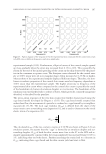
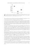
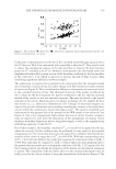
JOURNAL OF COSMETIC SCIENCE 186 inactivated cell extract (Sederma®, Sederma, Le Perray en Yvelines, France), prepared by freezing/thawing cycles. Cream containing 0.8 mM of CCSV (i.e., 0.8 mM phenylpro- panoids) was applied on skin surface a placebo cream was applied as control. After 5 days of a daily topical application, release of PGE-2, IL-6, and IL-8 was measured as men- tioned earlier, or explants were prepared for immunohistochemistry, being frozen and vertically sectioned (7–10 μm) using a cryostat (Leica® CM15105, Leica Biosystems Inc. Buffalo Grove, IL). Presence of Kallikrein-related peptidase 5 (KLK5), formerly known as stratum corneum tryptic enzyme (SCTE), a desquamation enzyme, which en- ables corneodesmosome division and corneocyte release to take place, was evaluated through immunolabelling performed using anti-SCTE antibody (Abcam®, Cambridge, MA). All studies were performed at 37°C in 5% CO2. Statistical analysis values were expressed as means ±SDM of the results for at least three experiments. Anova and Student t test were used for comparisons p values 0.05 were considered statistically signifi cant. IN-VIVO STUDIES Several studies were performed between March 2013 and December 2013 to establish the in vivo effi cacy of CCSV. Each individual study lasted 1 month. For each study and accord- ing to the protocol, volunteers applied by themselves a cream containing 0.8 mM CCSV and/or its vehicle. All the applications were performed on a daily basis in normal condi- tions of use. Each volunteer acted as his own control. Written informed consent was ob- tained from all participants. Medical control and noninvasive methods were used. Clinical studies comply with the latest recommendations of the World Medical Association (Dec- laration of Helsinki, 1964, and its successive updates) and with the French law 2004-806 Figure 1. NHS at passage 7 in NHS-7 medium. Left: 2 days after seeding right: 5 days. ×250. Figure 2. NHS in various culture media with 10% of FCS after 3 days: (A) DMEMc DI3, (B) DMEMc + EGF, (C) KSFM/BPE + EGF, (D) NHS7 Red Oil staining, ×500.
Purchased for the exclusive use of nofirst nolast (unknown)
From: SCC Media Library & Resource Center (library.scconline.org)