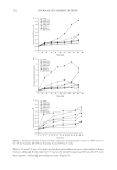
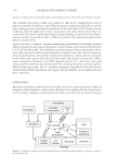
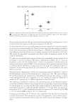
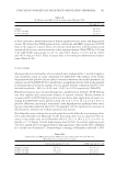
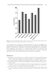
EVALUATION OF MOLECULES OR EXTRACTS MODULATING SEBORRHEA 189 Collagen-I coating, which helps NHK adhesion on plastic, did not modify signifi cantly neither anchorage of NHS, versus noncoated plates, nor proliferation. NHSs or NHDFs were embedded in collagen gels, respectively, in NHS-7 or in DMEMc both with 10% FCS, gel contraction was followed for 2 days. No contraction was ob- served with NHS (size reduction at day 2 vs. T0: 3%, nsd), whereas a quick and ex- pected contraction was observed with NHDF after 1 day (size reduction vs. T0: 52%, p 0.01). NHSs or NHKs were seeded on NHDF containing collagen lattice in KSFM + BPE and EGF media. Epidermis formation and differentiation were followed for 14 days using histology. No cell spreading was observed with NHS, whereas NHK produced as expected an epidermis with its stratum corneum. When NHSs seeded on collagen lattice were cultivated in NHS-7 medium, cells spread horizontally fi rst, then a multilayered “epidermis” was observed however, it was composed of small, rounded cells. No specifi c differentiation features were observed (i.e., neither spinous nor granular layers and no stratum corneum). Increase in size of NHS lipid droplets was observed using Red Oil staining depending on incubation time, inducers, serum concentration. NHS droplets are as numerous as in hu- man adipocytes but, conversely to adipocytes, they still remain small whatever the media used moreover, NHSs are smaller cells than adipocytes (Figure 4). As expected, lipid droplets are labelled with an anti-PLIN-2 antibody. Cells are positive for cytokeratin-7, and to a lesser extent for cytokeratin-4, which are, respectively, early and late differentiation markers of NHS (13,14). Rosiglitazone (0.1–10 μM), arachidonic acid (1–100 μM), linoleic acid (0.1–50 μM) all increased lipid storage into NHS from +44% to +665% (all p 0.01, vs. solvent). Conversely, CCSV (0.16–0.80 mM) re- duced lipid storage in a dose-dependent manner (p 0.01, vs. solvent Table I). NHS produces PGE-2, which can be enhanced in a dose-dependent manner with LPS (1.2–2.5 μg/ml Table II). CCSV (0.48 mM) was evaluated in parallel, showing an inhibi- tion of PGE-2 release without or with LPS. These inhibitions reached, respectively, 49% Table III PGE-2, IL-6, and IL-8 Release by NHK in Contact for 24 h with LPS. No Cell Toxicity Was Observed PGE-2 (pg/106 cell) IL-6 (pg/106 cell) IL-8 (pg/106 cell) No LPS 31.8 ± 4.3 2.6 ± 0.8 278 ± 53 LPS 1.2 μg/ml 111.6 ± 13.3 4.3 ± 0.2 368 ± 38 LPS 2.5 μg/ml 275.5 ± 88.4 3.9 ± 0.4 476 ± 52 LPS 5 μg/ml 402.5 ± 73.3 7.0 ± 0.5 861 ± 113 LPS 10 μg/ml 810.9 ± 112.6 33.2 ± 3.2 2483 ± 253 Table IV PGE-2, IL-6, and IL-8 Release by NHK in Contact with LPS and CCSV. No Cell Toxicity Was Observed PGE-2 (pg/106 cell) IL-6 (pg/106 cell) IL-8 (pg/106 cell) No LPS No CCSV 31.8 ± 4.3 2.6 ± 0.8 278 ± 53 CCSV 0.48 mM 23.1 ± 4.3 0,9 ± 0,13 121 ± 16 LPS 1.2 μg/ml No CCSV 111.6 ± 13.3 4.3 ± 0.2 368 ± 38 CCSV 0.48 mM 41.5 ± 5.5 1.68 ± 0.15 203 ± 15

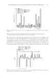
JOURNAL OF COSMETIC SCIENCE 190 and 47% (no LPS and 2.5 μg/ml LPS, both p 0.01). Similar results were observed with 0.16 and 0.8 mM CCSV. Results showed that proinfl ammatory mediators are produced by NHK in response to stress with a dose-dependent manner. NHK, in contact with LPS (1.2–10 μg/ml), re- leased PGE-2, IL-6, and IL-8 (Table III). PGE-2 release enhanced signifi cantly by 3.5× and 25.5×, whereas IL-6 release was triggered by 1.65× and 12.7×, and IL-8 release was signifi cantly enhanced by 1.3× and 8.9×, respectively, for 1.2 and 10 μg/ml. CCSV (0.48 mM) was evaluated in parallel without and with LPS it signifi cantly decreased the release of PGE-2, respectively, by -27% ( p 0.05) and 63% ( p 0.01), IL-6 -65% and -61%, (both p 0.01) and IL-8 by -57% and -45%, (both p 0.01) Table IV). Pro-infl ammatory mediators release was also studied on skin explants. Induction of PGE-2, IL-6, and IL-8 by skin explants was observed in contact with bacterial fragments. LPS and P. acnes fragments enhanced signifi cantly PGE-2 release by x7.4 and x7.1, IL-6 by x6.7 and x5.5, and IL-8 by x23.9 and x7.5 (Table V). CCSV (0.8 mM) was evaluated in paral- lel it inhibited the strong enhancement of PGE2 due to LPS or P. acnes fragments and maintained these release to their control level, same observation was made for IL-6, CCSV stabilizing the cytokine production to a basal level. For IL-8 release, CCSV was prone to regulate more effi ciently its production: by -90% under LPS stimulus and by -34% with P. acnes (both p 0.01 Table VI). These results, obtained with NHS, NHK, and explants confi rmed that bacterial frag- ments both from E. coli and P. acnes triggered proinfl ammatory mediators and indicated that CCSV controls their stress increase production. Since Al Shobaili et al. (15) did show a correlation with acne vulgaris and oxidative and nitrosative stress, a study was performed on RAW 264.8 activated with LPS, in order to evaluate the control of NO release owing to CCSV. Results (Table VII) showed a dose- dependent decrease of NO release by -41% and -56% (both p 0.01), respectively, with 0.16 and 0.48 mM CCSV. It was shown that sebaceous glands and sebocytes participate to innate immunity by pro- ducing AMP such as β-defensin-2 and cathelicidin (16,17). AMPs were studied on NHS Table V PGE-2, IL-6, and IL-8 Release by Skin Explants in Contact for 24 h with LPS or P. acnes PGE-2 (pg/ml) IL-6 (pg/ml) IL-8 (pg/ml) Control 220 ± 40 442 ± 30 278 ± 69 LPS 5 μg/ml 1635 ± 280 3038 ± 872 6650 ± 1019 P. acnes fragments 1569 ± 523 2419 ± 752 2079 ± 820 Table VI PGE-2, IL-6, and IL-8 Release by Skin Explants in Contact with LPS and CCSV PGE-2 (pg/ml) IL-6 (pg/ml) IL-8 (pg/ml) LPS 5 μg/ml Placebo cream 1635 ± 280 3038 ± 872 6650 ± 1019 Cream with CCSV 0.8 mM 117 ± 33 390 ± 231 659 ± 259 P. acnes Placebo cream 1569 ± 523 2419 ± 752 2079 ± 820 Cream with CCSV 0.8 mM 171 ± 48 427 ± 216 1392 ± 518
Purchased for the exclusive use of nofirst nolast (unknown)
From: SCC Media Library & Resource Center (library.scconline.org)