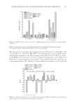
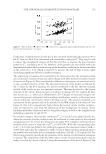
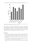
EVALUATION OF MOLECULES OR EXTRACTS MODULATING SEBORRHEA 193 acid, arachidonic acid, rosiglitazone, and CCSV. Moreover, the release of proinfl ammatory mediators under various external stresses (lipids and bacterial fragments) could be easily followed and results were compared to data obtained with keratinocytes, macrophages, both neighboring cells of sebocytes, or explants models. CCSV was studied here with various skin-derived cell models and in vivo results indicated its interest as a bioactive ingredient for the control of hyperseborrhea and its negative consequences. REFERENCES (1) M. Dalhoff, E. Camera, M. Picardo, C. C. Zouboulis, L. Chan, B. H. Chang, and M. R. Schneider, PLIN2, the major perilipin regulated during sebocyte differentiation, controls sebaceous lipid accumu- lation in vitro and sebaceous gland size in vivo., Biochem. Biophys. Acta., 1830, 4642–4649 (2013). (2) Ottaviani M., Camera E., and Picardo M., Lipid Mediators in Acne, Mediat. Infl amm., 2010, 1–6 (2010). (3) M. Picardo, M. Ottaviani, E. Camera, and A. Mastrofrancesco, Sebaceous gland lipids, Dermato-Endroc., 1, 68–71 (2009). (4) N. Akimoto, T. Sato, C. Iwata, M. Koshizuka, F. Shibata, A. Nagai, M. Sumida, and A. Ito, Expression of perilipin A on the surface of lipid droplets increases along with the differentiation of hamster sebo- cytes in vivo and in vitro, J. Invest. Dermatol., 124, 1127–1133 (2005). (5) B. T. Toth, A. Olah, A. G. Szollosi, G. Czifra, and T. Biro, Sebocyte makeup: Novel mechanism and concepts in the physiology of the human sebaceous glands, Pfl ugers Arch-Eur J. Physiol., 461, 593–606 (2011). Figure 6. Percent of satisfi ed opinions after 1 month, n = 100 vol. cream with CCSV 0.8 mM. All bars indicate a signifi cant variation versus negative opinions with p 0.01 (χ2 test).

JOURNAL OF COSMETIC SCIENCE 194 (6) D. Thiboutot, S. Jabara, J. McAllister, A. Sivarajah, K. Gilliland, Z. Cong, and G. Clawson, Human skin is a steroidogenic tissue: steroidogenic enzymes and cofactors are expressed in epidermis, normal sebocytes, and an immortalized sebocyte cell line (SEB-1), J. Invest. Dermatol., 120, 905–914 (2003). (7) C. Zouboulis, C. H. Seltmann, H. Neitzel, and C. E. Orfanos, Establishment and characterization of an immortalized human sebaceous gland cell line (SZ95), J. Invest. Dermatol., 113, 1011–1020 (1999). (8) P. Greenspan, E. Mayer, and S. D. Fowler, Nile red: A selective fl uorescent stain for intracellular lipid droplets, J. Cell Biol., 100, 965–973 (1985). (9) E. Bell, B. Ivarsson, and C. Merrill, Production of a tissue-like structure by contraction of collagen lat- tices by human fi broblasts of different proliferative potential in vitro, Proc. Natl. Acad. Sci. U.S.A., 76, 1274–1278 (1979). (10) M. W. Carlson, A. Alt-Holland, G. Egles, and J. A. Garlick, Three dimensional tissue models of normal and diseased skin, Curr. Protoc. in Cell Biol., Chapter Unit:19.9, Vol. 41,19.9 .1-19.9.17. (2008) (11) J. Kim, M. T. Ochoa, S. R. Krutznik, O. Takeuchi, S. Uematsu, A. J. Legaspi, H. D. Brightbill, D. Holland, W. J. Cunliffe, S. Akira, P. A. Sieling, P. J. Godowski, R. L. Modlin, Activation of toll-like receptor 2 in acne triggers infl ammatory cytokine responses, J. Immunol., 169, 1535–1541 (2002). (12) W. J. Lee, K. H. Park, H. W. Cha, M. Y. Sohn, K. D. Park, S. J. Lee, D. W. Kim, The expression of involucrin, loricrin and fi laggrin in cultured sebocytes., Ann. Dermatol., 26, 134–137 (2014). (13) E. Hinde, I. S. Haslam, M. R. Schneider, E. A. Langan, J. E. Kloepper, C. Schramm, C. C. Zouboulis, and R. Paus, A practical guide for the study of human and murine sebaceous glands in situ, Exp. Dermat., 22, 631–637 (2013). (14) C. C Zouboulis, L. Xia, B. Korge, H. Gollnick, and C. E. Orfanos, “Cultivation of human sebocytes in vitro: Cell characterization and infl uence of synthetic retinoids,” in Retinoids: 10 Years On, J. H. Saurat. Ed. (Karger, Basel, 1991), pp. 254–273. (15) H. A. Al-Shobaili, A. A. Alzolibani, A. A. Robaee, A. R. Meki, and Z. Rasheed, Biochemical markers of oxidative and nitrosative stress in acne vulgaris: Correlation with disease activity, J. Clin. Lab. Anal., 27, 45–52 (2013). (16) D. Y. Lee, K. Yamasaki, J. Rudsil, C. C. Zouboulis, G. T. Park, J. M. Yang, and R. L. Gallo, Sebocytes express functional cathelicidin antimicrobial peptides and can act to kill propionibacterium acnes, J. Invest. Dermatol., 128, 1863–1866 (2008). (17) M. R. Benakanakere, Q. Li, A. V. Singh, J. Zhao, and D. F. Kinane, Modulation of TLR2 protein expres- sion by miR-105 in human oral keratinocytes, J. Invest. Dermatol., 284, 23107–23115 (2009).
Purchased for the exclusive use of nofirst nolast (unknown)
From: SCC Media Library & Resource Center (library.scconline.org)