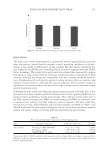
J. Cosmet. Sci., 68, 195–204 (May/June 2017) 195 Noninvasive measurement of advanced glycation end-products in the facial skin: New data for skin aging studies DI QU, DAWNA VENZON, MARY MURRAY, and MATHEW DEPAUW Amway R&D, Ada, MI. Accepted for publication April 10, 2017. Synopsis Using skin autofl uorescence (SAF) as a marker of advanced glycation end-products (AGEs) has been extensively studied in the last decade since the introduction of the noninvasive in vivo measurement technique. Data have shown the level of skin AGEs increases with chronological age in healthy human beings, and this increase is substantially higher in age-matched diabetic patients. In skin research, glycation with the accompanying accumulation of skin AGEs has been regarded as one of the primary skin aging mechanisms that contribute to skin wrinkling and the loss of skin elasticity. To date, the totality of SAF data reported in literature has been obtained from measurements on the arm, and noninvasive measurement of facial skin AGE accumulation would add great value to skin aging research. In this study, we report the levels of facial and forearm skin AGEs in 239 men and women of 21–65 year of age. Signifi cantly lower levels of AGEs were detected in the facial skin than in the forearm skin from the young Caucasian groups, and the difference was much larger for men than for women. The rate of change in skin AGE level over age was found to be about 50% higher in men than in women, which further highlights the gender difference. A statistically signifi cant correlation between the levels of skin AGE and facial wrinkling was also observed. The facial skin AGE data may provide new insight into skin aging research. INTRODUCTION Skin autofl uorescence (SAF) has been well validated in the past 10 years after the fi rst paper appeared in the literature describing a simple noninvasive method to measure the accu- mulation of the advanced glycation end-products (AGEs) in the skin (1). AGEs are formed in the tissue via a nonenzymatic glycation process between sugars and proteins and are implicated in the pathophysiology of aging, including complications of multiple aging- related diseases (2–5). With the convenience of the noninvasive measuring technique, skin AGEs have been measured in large study populations. As such, SAF has been estab- lished as a noninvasive marker of vascular damage in Caucasians patients with type 2 diabetes (6–10), as a strong predictor of cardiac mortality in diabetes (11), as a comple- mentary test to assess kidney function as well as to predict mortality in hemodialysis patients (12–14), and as a novel risk marker in chronic kidney disease (15). In addition to Address all correspondence to Di Qu at di.qu@amway.com.

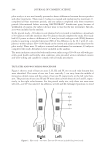
JOURNAL OF COSMETIC SCIENCE 196 diabetic and renal diseases, elevated levels of skin AGEs have been reported in patients with systemic lupus erythematosus (16), chronic cerebral ischemia (17), schizophrenia (18), peripheral artery disease (19,20), and chronic heart failure (21). SAF has also been studied in healthy people, and positive correlations of skin AGE levels with chronological age have been established for Dutch and Slovak Caucasians, Japanese, Chinese, and Saudi Arabians (22–26). Because skin pigmentation can infl uence the results of SAF, corrections for melanin and hemoglobin exist to enable more meaningful correla- tions with age in those individuals with darker skin pigments (27). In the skin, AGEs are considered photosensitizers and can generate reactive oxidative species on ultraviolet (UV) irradiation, which accelerates the skin aging process (28,29). Meanwhile, there have been reports indicating that chronic UVB exposure induces additional fl uorescence exci- tation bands in mice skin (30) and observations of signifi cantly more AGE staining in sun-exposed skin than in sun-protected skin, suggesting that solar irradiation increases dermal glycation (31). Increased skin AGEs has also been correlated with heavy smokers and chronic obstructive pulmonary disease patients (32). Of the SAF studies, most of them has used commercially available noninvasive instru- ments, AGE Reader (DiagnOptics, Groningen, The Netherlands) or SCOUT DS (VeraLight, Albuquerque, NM), to measure the accumulated skin AGEs in vivo. Their detailed oper- ating principles were previously reported (1,8). To date, all studies using SAF as a mea- sure of AGEs have been obtained from the volar forearms except two reports in which the measurements were obtained on the inner aspect of the upper arm skin (23,33). The facial skin has been the primary focus of antiaging research in the skincare industry, and the noninvasive in vivo measurement of facial skin AGEs would add new data to ag- ing research. In this study, we report skin AGEs measured from the left-cheek skin and compare the results with that of the left volar forearm skin. Our aim was to show the site and gender differences of skin AGE level and to correlate the results with participants’ chronological age as well as the level of facial wrinkling. We selected the left cheek for measurement to maximize the effect of sun exposure on the skin because of the driving convention in the United States. MATERIALS AND METHODS SAF MEASUREMENT A commercially available AGE Reader™ SU (DiagnOptics) was used to noninvasively evaluate the level of accumulated AGEs in the skin. Its measurement principle is based on the properties of SAF because the primary components of AGEs in the skin emit a characteristic SAF when excited by UV light. The instrument illuminates a skin surface area of 4 cm2, guarded against surrounding light, with an excitation light source between 300 and 420 nm (peak excitation 370 nm). Emission light and refl ected excitation light from the skin are measured with a spectrometer in the 420–600 nm range. Because skin pigmentation may absorb light and thus infl uence autofl uorescence, skin refl ection mea- surements across the 300–420 nm range were compared with those of a white Tefl on block (1). SAF is calculated as the ratio of the light intensity refl ected by the skin in the 420–600 nm wavelength range and the light intensity in the 300–420 nm wavelength range and is represented as the skin autofl uorescence ratio (AFR) in AGE Reader (1).
Purchased for the exclusive use of nofirst nolast (unknown)
From: SCC Media Library & Resource Center (library.scconline.org)